
Translate this page into:
Management of Babesiosis in a Nigerian Local Breed of Dog: A Clinical Case Study

*Corresponding author: Amaka Rosita Okonkwo, Department of Biochemistry and Drug Development, National Veterinary Research Institute, Jos, Nigeria. akpaamaka@gmail.com
-
Received: ,
Accepted: ,
How to cite this article: Okonkwo AR, Uchendu C, Idoga ES, Gurumyen GY, Madubueze JS. Management of Babesiosis in a Nigerian Local Breed of Dog: A Clinical Case Study. Res Vet Sci Med. 2025;5:2. doi: 10.25259/RVSM_12_2024
Abstract
Canine Babesiosis is a worldwide tick-borne disease. In dogs, it is usually caused by Babesia canis. On physical examination, the dog was febrile, it had a high pulse and respiratory rates, the submandibular lymph nodes were bilaterally enlarged, the ocular mucus membranes were pale, there was heavy tick infestation, dark-colored urine, the perineum was matted, and the dog was weak. Peripheral blood smear examination revealed the presence of Babesia parasites in the erythrocytes, severe anemia, icterus, neutrophilia, anisocytosis, macrocytosis, and polychromatophilia. This case is an uncomplicated case of babesiosis and the dog recovered after the treatment regimen. This study aims to report a case of babesiosis in a Nigerian Local dog, describe the clinical presentations, briefly describe the pathophysiology of the clinical signs seen, and illustrate the importance of early diagnosis and rational treatment strategies.
Keywords
Babesiosis
Malignant jaundice
Nigerian local dog
Regenerative anemia

INTRODUCTION
Canine Babesiosis also called “malignant jaundice” or “tick fever” is a potentially fatal and a major disease of dogs in Nigeria.[1] It is a tick-borne protozoan infection caused majorly by two Babesia species, Babesia canis and Babesia gibsoni in dogs. Babesia spp. is an intracellular apicomplexan protozoan parasite that infects and replicates in the red blood cells of dogs causing anemia.[2] It is primarily transmitted by the brown dog tick (Rhipicephalus sanguineus) and is one of the most common piroplasmic infections of dogs.[3] A tick carrying B. canis sporozoites attaches to a dog and feeds on its blood, releasing many sporozoites into the dog’s bloodstream.[1,2] Each sporozoite attaches to a red blood cell and moves inside the cell. Once inside the cell, the sporozoite loses its outer coating and divides, becoming a new form, known as a merozoite.[3] Inside the tick, the merozoite undergoes sexual reproduction (gamogony), which is followed by asexual reproduction, resulting in many sporozoites.[3] These are found in the tick salivary glands, and they move from there into the next dog on which the tick feeds.[4] In most cases, B. canis is spread to dogs through the bite of an infected tick;[5] however, some studies suggest that infected dogs with open-mouth sores can pass on the infection to other dogs through a bite,[6] and infected pregnant females can transmit babesiosis to their unborn puppies.[7] Canine babesiosis can be clinically classified into uncomplicated and complicated forms. Clinical signs seen in the uncomplicated form of babesiosis are suggested to be a consequence of hemolysis whereas complicated canine babesiosis has been suggested to be a consequence of the development of systemic inflammatory response, secondary bacterial infections, and multiple organ dysfunction syndrome.[8] The purpose of this case report is to investigate a clinical case of Babesiosis in a Nigerian Local Dog, with clinical and laboratory examinations performed, and to advise on preventive measures.
CASE REPORT
A 10-month-old male Nigerian local dog weighing 11.3 kg was presented on June 6th, 2024 to the small animal clinic of the University of Jos Veterinary Teaching Hospital with the chief complaint of anorexia, which has lasted for 2 days.
On physical examination, the dog was febrile (40.2°C), had a slightly high pulse rate (148 beats/min), a slightly high respiratory rate (35 cycles/min), and bilaterally enlarged submandibular lymph nodes. Further clinical examination revealed a severely pale ocular mucus membrane [Figure 1], epilation, heavy tick infestation, lethargy, depression, dark-colored urine, and a matted perineum.
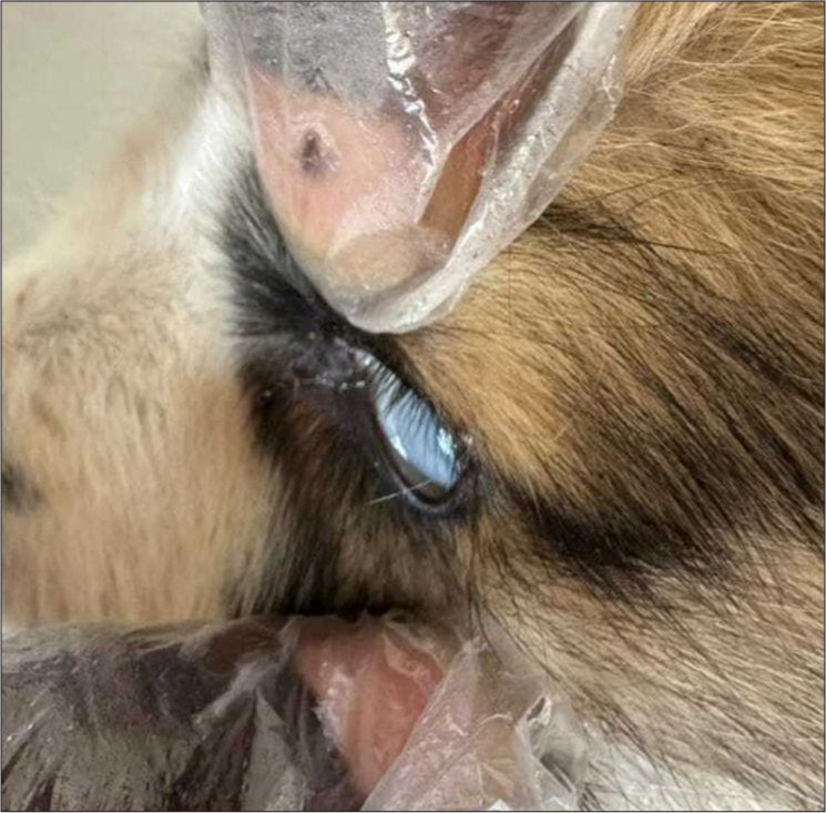
- Picture of an anemic Nigerian local dog shows a very pale ocular mucus membrane.
Peripheral blood was collected in a sample bottle containing ethylenediamine tetraacetic acid for a blood smear examination and hematology. Blood smear examination revealed the presence of pear-shaped piroplasmic organisms in the erythrocytes [Figure 2], while hematology revealed a very severe anemia (packed cell volume of 13%), jaundice (yellow colored plasma), neutrophilia, anisocytosis, macrocytosis, and polychromatophilia indicative of severe regenerative anemia. The tick infesting the dog was identified as a Rhipicephalus species.
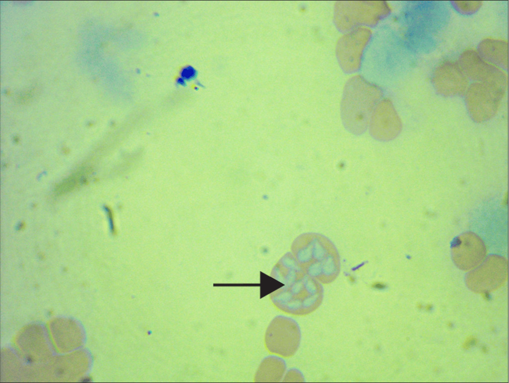
- Photomicrograph of the infected patient. The black arrow shows red blood cells infected with pear-shaped Babesia spp.
DISCUSSION
Based on clinical and laboratory findings, the condition was diagnosed as babesiosis. An initial treatment with proxicam at 0.3 mg/kg intramuscularly and B-complex vitamins was administered on presentation and after confirmation, imidocab dipropionate was administered at a dose of 6.6 mg/kg subcutaneously once, 1% ivermectin at 0.6 mg/kg subcutaneously once to treat the heavy tick infestation, piroxicam at 0.3 mg/kg intramuscularly for 2 days for pain management, oxytetracycline long acting at 20 mg/kg intramuscularly once for secondary bacterial infections, and iron dextrans 10 mg/kg intramuscularly once to stimulate erythropoiesis. The dog recuperated progressively, with full recovery noted 2 weeks after treatment.
Babesia spp. are intracellular parasitic organisms that affect mainly the red blood cells of most mammals, causing babesiosis.[7] Babesiosis is caused by both the large and small forms of Babesia species (B. canis, Babesia vogeli, B. gibsoni, and Babesia microti) in dogs and is usually transmitted by ticks.[9] Babesiosis is potentially fatal and a major economically important disease of dogs and this disease is endemic in Nigeria.[10] A study by Obeta et al.[11] revealed that this parasite was identified in apparently healthy dogs in the federal capital territory of Nigeria with the disease being more prevalent during the rainy season than during the dry season and older dogs being more resistant than younger dogs.[11]
Dogs with uncomplicated babesiosis such as the above case usually exhibit clinical signs of fever, anorexia, lethargy, and depression, and these findings are in accordance with previous research works by Mahdy et al.[12] The clinical signs observed in the present case include signs of depression such as weakness, restlessness and anorexia, tachycardia, tachypnea, and fever. It is thought that the clinical signs are the result of tissue hypoxia caused by severe anemia and concomitant systemic inflammatory response syndrome which was caused by marked cytokine release.[12] Anemia seen in this case was a result of excessive destruction of red blood cells in the spleen and liver by erythrophagocytosis. Furthermore, immunoglobulin and complement-mediated destruction of both parasitized and non-parasitized erythrocytes are an important aspect of anemia and previous studies have shown a correlation between anti-erythrocyte antibodies and anemia in B. gibsoni infection.[13] Icterus is caused by the presence of high concentrations of conjugated bilirubin and caused by inactivation of the transfer system for conjugated bilirubin between hepatocytes and bile canaliculi.[14] The degree of functional impairment, hepatocellular damage, and bile stasis in canine babesiosis is indicative of the degree of icterus observed.[15]
Imidocarb dipropionate was the antiprotozoan agent used for treatment in this case. It is an aromatic diamidine, which acts by blocking the entry of inositol into erythrocytes containing Babesia spp. This results in starvation of the parasite, interference with production and/or use of polyamines by parasites, nucleic acid damage, and inhibition of cellular repair and replication.[6] Vitamin B complex was administered as a supplement for the nutritional rehabilitation necessary for tissue repair in an acute febrile systemic disease in which a degree of hepatopathy frequently co-exists and in which the hepatic storage of vitamins could be deficient. Antibiotics in the form of oxytetracycline long-acting were administered to avoid or minimize any secondary or opportunistic bacterial infection, providing a bacteriostatic antibiotic cover in a patient whose reticuloendothelial system may be overburdened by the insults of the protozoan disease. Piroxicam is a non-steroidal anti-inflammatory drug which was administered in this case to combat pyrexia and inflammatory conditions seen in this case. Piroxicam acts by inhibiting the cyclooxygenases 1 and 2 enzymes which are responsible for producing proinflammatory prostaglandins which are mediators of pain and inflammation.[16] Ivermectin is an antiparasitic drug. It was administered in this case to treat the tick infestation. Ivermectin acts by binding to the chloride ion channels of nerve or muscle cells immobilizing the tick inducing tonic paralysis of the buccal musculature and other muscles in the tick.[17]
We advised the client to use acaricides such as cypermethrin spray to control ticks on other dogs and in the environment so as to prevent reinfestation of the dogs with ticks.
CONCLUSION
In the present case, combination therapy using imidocarb dipropionate and oxytetracycline along with the other drugs used in supportive care was effective in ameliorating the clinical signs of Babesia infection, eliminating parasitemia and aiding in early recovery of the animal. However, these hemoparasites are seldom completely eliminated, and when immunocompromised, reinfection may occur. Hence, we advised the client on the need to regularly control ticks to prevent the recurrence and spreading of infection. This can be achieved by regular tick bathing of dogs, cleaning of surrounding bushes, and regular washing and spraying of kennels with acaricides. Dog breeders and keepers should ensure trimming of bushes and grasses where ticks are a serious problem. A dirty, wet, and moist environment which enhances the survival of the tick’s vectors that transmitted the babesiosis should be controlled and avoided, and report to the closest veterinarian when he notices any form of tick infestations or illnesses rather than self-medicate.
Acknowledgments
The author acknowledges the contributions of Dr. Paul for his assistance.
Ethical approval
Institutional Review Board approval is not required.
Declaration of patient consent
Patient’s consent not required as there are no patients in this study.
Conflicts of interests
There are no conflicts of interest.
Use of artificial intelligence (AI)-assisted technology for manuscript preparation
The authors confirm that there was no use of artificial intelligence (AI)-assisted technology for assisting in the writing or editing of the manuscript and no images were manipulated using AI.
Financial support and sponsorship: Nil.
References
- Prevalence of Babesia Spp. In Presumably Healthy Dogs and Associated Risk Fators in OBIO/AKPOR Local Government Area. Rivers State, Nigeria. J Appl Vet Sci. 2023;8:89-97.
- [CrossRef] [Google Scholar]
- Pathogenesis of Anemia in Canine Babesiosis: Possible Contribution of Pro-inflammatory Cytokines and Chemokines-A Review. Pathogens. 2023;12:166.
- [CrossRef] [PubMed] [Google Scholar]
- Parasites in the Cardiovascular System In: Organ-specific Parasitic Diseases of Dogs and Cats. United States: Academic Press; 2023. p. :53-88.
- [CrossRef] [Google Scholar]
- Protozoan Infections in Dogs and Cats In: Principles and Practices of Canine and Feline Clinical Parasitic Diseases. United States: John Wiley and Sons; 2024. p. :119-31.
- [CrossRef] [Google Scholar]
- Diagnosis and Treatment of Canine Babesiosis in a Female German Shepherd in Tandojam, Pakistan. Mathews J Vet Sci. 2024;8:44.
- [CrossRef] [Google Scholar]
- The Etiology, Incidence, Pathogenesis, Diagnostics, and Treatment of Canine Babesiosis Caused by Babesia gibsoni Infection. Animals (Basel). 2022;12:739.
- [CrossRef] [PubMed] [Google Scholar]
- Histopathological Aspects of the Influence of Babesia microti on the Placentas of Infected Female Rats. Vet Sci. 2024;11:18.
- [CrossRef] [PubMed] [Google Scholar]
- Studies on the Pathophysiology of Babesia gibsoni Infection in Pakistani Bully, Shih TZU, German Shepherd and Labrador Dogs. Int J Vet Sci Anim Husb. 2024;9:582-5.
- [Google Scholar]
- Microscopical and Molecular Diagnosis of Canine Babesiosis in Stray Dogs in Erbil, Iraq. Iraqi J Vet Sci. 2024;38:823-30.
- [CrossRef] [Google Scholar]
- Genetic Characterization of the RAP-1A and SBP-4 Genes of Babesia Species Infecting Cattle from Selangor, Malaysia, and Ribah, Nigeria. Pathogens. 2024;13:247.
- [CrossRef] [PubMed] [Google Scholar]
- Prevalence of Canine Babesiosis and their Risk Factors among Asymptomatic Dogs in the Federal Capital Territory, Abuja, Nigeria. Parasite Epidemiol Control. 2020;11:e00186.
- [CrossRef] [PubMed] [Google Scholar]
- Genetic Characterization and Pathogenic Effects of Hepatozoon canis Infection in Police Dogs in Egypt. Beni Suef Univ J Basic Appl Sci. 2024;13:40.
- [CrossRef] [Google Scholar]
- Lessons Learned for Pathogenesis, Immunology, and Disease of Erythrocytic Parasites: Plasmodium and Babesia. Front Cell Infect Microbiol. 2021;11:685239.
- [CrossRef] [PubMed] [Google Scholar]
- Feline Cytauxzoonosis: Reconsideration of Pathophysiologic Mechanisms (Doctoral Dissertation) In: Kansas State University. 2022.
- [Google Scholar]
- Differential Diagnoses in 83 Dogs with Icterus. Pesqui Vet Bras. 2020;40:451-65.
- [CrossRef] [Google Scholar]
- Insight Into the Mechanism of Steroidal and Non-steroidal Anti-inflammatory Drugs In: How Synthetic Drugs Work. United States: Academic Press; 2023. p. :61-94.
- [CrossRef] [Google Scholar]
- Ivermectin: An Anthelmintic, An Insecticide, and Much More. Trends Parasitol. 2021;37:48-64.
- [CrossRef] [PubMed] [Google Scholar]


