
Translate this page into:
Management of Cubital Joint Re-luxation Using a Definitive Approach of Transarticular External Skeletal Fixator

*Corresponding author: Abdulhakeem Binhambali, Department of Clinical Sciences, College of Veterinary Medicine, Tuskegee University, Tuskegee, United States. email2hambali@gmail.com
-
Received: ,
Accepted: ,
How to cite this article: Bada AA, Lanko ZS, Binhambali A, Bappah MN, Gaba EE. Management of Cubital Joint Re-luxation Using a Definitive Approach of Transarticular External Skeletal Fixator. Res Vet Sci Med. 2025;5:7. doi: 10.25259/RVSM_1_2025
Abstract
This is a case of a 6-month-old Alsatian cross female puppy diagnosed with cubital joint re-luxation. The puppy presented to the clinic with a complaint of an inability to walk with the right forelimb following an initial surgery to address a cubital luxation that was due to a road traffic accident. Radiographic imaging confirmed the luxation of the cubital joint, which was managed with an initial open reduction using cerclage wiring. However, the postoperative evaluation revealed joint re-luxation. The patient was managed with a definitive surgical approach involving transarticular external skeletal fixation. The case report provides a detailed description of the clinical history, diagnostic findings, surgical procedures, and postoperative management of this case. The patient came back with minor lameness post 1-year follow-up, which made the outcome a successful approach.
Keywords
External skeletal fixative
Fractures
Joint luxation
Transarticular pin

INTRODUCTION
Cubital joint luxation (CL) is a common orthopedic condition in dogs which is majorly caused by either trauma[1] or idiopathic congenital development.[2] Neither congenital nor early acquired elbow luxation through trauma or traffic accidents are common clinical presentations in dogs,[3] and these can be encountered in both males and females and may affect one or both sides of the elbow.[4] There is limited documentation regarding the treatment strategies for such cases, especially re-luxation after previous management in existing literature due to poor response to treatment or management from the initial surgery.[2]
Arthrodesis, a surgical technique in veterinary medicine, involves permanently immobilizing a joint to address issues like severe instability or chronic pain that is not responsive to other treatments.[5] It is sometimes necessary for puppies to correct congenital abnormalities or injuries that affect joint function.[6] The outcome of arthrodesis in puppies depends on factors such as their age during presentation, the specific joint involved, the underlying condition, and the surgical approach used. Temporary arthrodesis may be considered in cases where there’s potential for a joint function to recover after a period of immobilization.[7] This approach stabilizes the joint temporarily while allowing for the possibility of future mobility. However, the success of this temporary fusion varies based on individual factors and the underlying condition being treated, as stated earlier. Permanent arthrodesis, on the other hand, involves the complete fusion of the joint surfaces, resulting in permanent immobilization. While this approach effectively stabilizes the joint, it also eliminates any potential for joint motion. Therefore, the restoration of joint function following permanent arthrodesis in puppies is typically not expected.[8]
The major cause of CL in a puppy dog is often attributed to trauma or injury, particularly during periods of rapid growth when the bones and joints are still developing and vulnerable to damage[9], and management of such cases requires careful diagnostic evaluation and appropriate surgical intervention to restore joint function and prevent long-term complications.
Cubital re-luxation in dogs is the abnormal displacement of the elbow joint (cubital joint) after a previous dislocation, often due to ligamentous instability or inadequate stabilization post-injury. It commonly results from trauma, congenital defects, or surgical complications. Affected dogs may exhibit obvious lameness, pain, joint swelling, and reduced range of motion. Diagnosis involves radiographs or advanced imaging, while treatment may include splinting, surgical stabilization, or joint reconstruction, depending on severity. In this paper, we report the management of a cubital re-luxation using a successful surgical procedure involving the use of trans-articular external skeletal fixation.
CASE REPORT
An Alsatian cross female intact 6 months puppy (16.5 kg) was referred to Ahmadu Bello University Veterinary Teaching Hospital, small animal surgery unit on the 26th of July, 2021, and was examined for a mobility problem and inability to walk specifically with the right forelimb. The referral letter revealed that the initial problem was noticed a month earlier before the presentation. History also revealed that the puppy was hit by a motorcycle when the owner was on a road walk with the puppy. On examination, the right forelimb was shorter than the normal contra-lateral forelimb and was arched with greater pronation of the distal part of the forelimbs. There was also slight medial concavity of the antebrachia just below the elbow joint. Following radiographic imaging of the right forelimb, luxation of the cubital joint was diagnosed [Figure 1a]. The condition was initially managed by open reduction using cerclage wiring only [Figure 1b]. However, three weeks after the initial management, post-operative radiographic imaging revealed the re-luxation of the cubital joint [Figure 1c], and this postoperative revelation necessitated a definitive surgical procedure involving the use of external skeletal fixation.
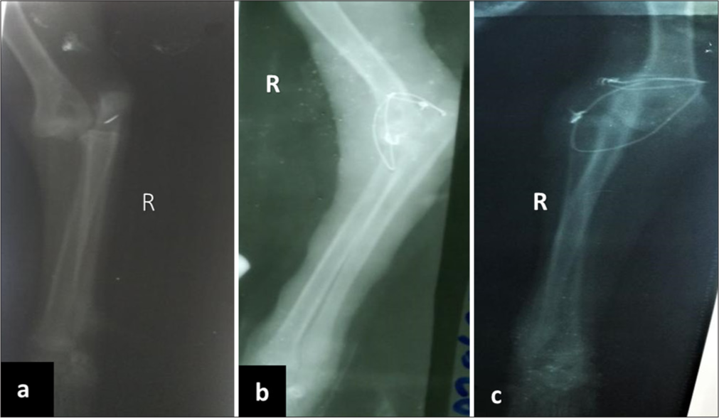
- (a) Mediolateral view of the right forelimb showing medial displacement of the humerus on presentation; (b,c) Radiographs showing re-luxation of the cubital joint three weeks after the initial management.
Case management
The patient vital parameters on the 1st day of presentation were presented in Table 1. The patient was induced with the pre-anesthetic protocol of injectable atropine sulfate 0.02 mg/kg (0.33 mg, Amopin®, YanzhouXierkangtaiPharma Ltd, China) intravenous (I.V) once, Chlorpromazine 2 mg/ kg (33 mg, Pauco Chlorpromazine hydrochloride (HCL) Injection®, Pauco Pharmaceutical Ltd, Nigeria) I.V once. The surgical site was shaved to expose the underlying skin of the right shoulder joint to the level of the carpal joint and the site was scrubbed thoroughly with 0.05% Chlorhexidine gluconate (Purit®Saro Lifecare Ltd, Ibadan, Nigeria) and Povidone iodine solution (Aqua Poviderm®Aqua Medical Ltd., Istanbul) to remove any underlying dirt and to maintain asepsis during the time of surgery. Anesthesia was induced using injectable thiopentone sodium 10 mg/kg (165 mg, Thosol sodium, Neon Laboratories Ltd, Mumbai India) I.V. and maintained with ketamine hydrochloride 10 mg/kg (165 mg, Pauco Ketamine Injection®, Kwality Pharmaceutical Ltd, India) I.V as available in the country. The surgical site was draped and clamped with a Backhaus towel clamp in a triangular format, exposing only the site of surgical interest.
| Parameters | Patient values | Reference values |
|---|---|---|
| Temperature (°C) | 38.8 | 38.5–39.4 |
| Pulse rate (beats/min) | 102 | 90–120 |
| Respiratory rate (cycles/min) | 30 | 20–30 |
As described by Campbell,[10] a skin incision was made on the lateral surface of the elbow immediately over the lateral prominence of the lateral epicondyle of the humerus. Blunt dissection between the extensor muscles of the carpus, which have their origin in the lateral epicondyle, allows the insertion of a lever over the head of the radius and under the lateral epicondyle of the humerus. The cerclage wires from the first surgical procedure were removed, and the joint was separated. Using downward pressure on this lever, the joint surfaces were forced apart a little and then, by lateral pressure, returned to normal position [Figure 2].
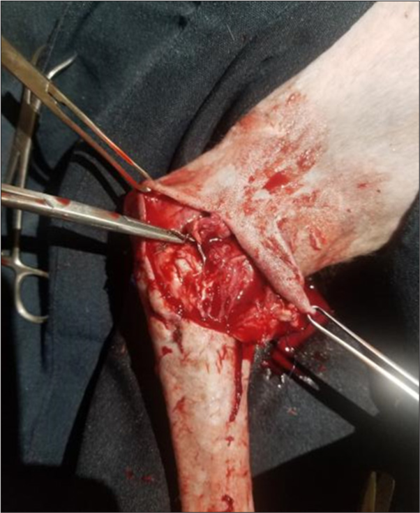
- Dissection through the muscles, exposure, and evaluation of the fracture site on the cubital joint.
Afterward, skin stab incisions were made on the lateral side of the skin of the arm (humerus) and forearm (radius), where the intended transfixion pins will be drilled into [Figure 3]. The transfixion pins (Front Threaded Pin, Shanz Screw 3.5 × 200 mm, stainless steel, Asco, India) were drilled through the stab incision site into the humerus, radius, and ulna bones. Three pins were inserted into the humerus: one each into the proximal, midshaft, and distal humerus and one pin each into the olecranon process, proximal, midshaft, and distal radius. The three pins in the humerus were connected to three clamps (Small Single Pin Clamp 4.0 × 3.5 mm, Asco, India) and a single connecting bar (Connecting Rod 4.0 mm × 300 mm, Asco, India), while the four pins in the radius and olecranon process were similarly connected to a single connecting bar using four clamps. The two connecting bars were connected using a single clamp at the level of the elbow which serves as a hinge (joints) between the humerus and the radius-ulna [Figure 4]. All the clamps were adequately tightened using a spanner except the clamp holding the two connecting bars together. The elbow joint was put at a proper standing angular position similar to the contra-lateral forelimb with the aid of a goniometer. Finally, the clamp holding the two connecting bars were adequately tightened, and the elbow incision was closed routinely with intradermal suture.
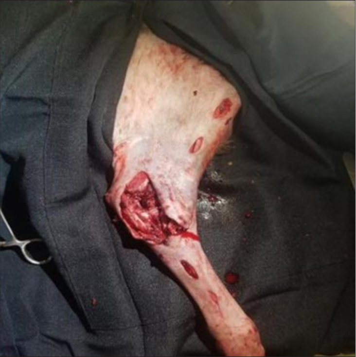
- Incisions were made at points to be drilled on the humerus and radio-ulnar to expose bones for drilling.

- (a,b) show the placement of the External Skeletal Fixator which extends from the proximal humerus to the distal radio-ulnar.
The surgical site was thoroughly cleaned before the bandage was placed on it [Figure 5]. Post-surgical management was done using Injectable Piroxicam 0.3 mg/kg (4.95 mg, Philoxicam®, Tianjin Kingyork Hubei TianyaoPharm. Co. Ltd, Hubei, China) I.M o.d × 7/7 to manage pain, and injectable Ceftriaxone 30 mg/kg (495 mg, Eden Ceftriaxone®, Eden U.K Pharmaceuticals Ltd, Anambra, Nigeria ) intramuscular (I.M) o.d × 7/7 to manage post-surgical infection. The patient’s vital parameters were monitored on subsequent days post-surgery, as listed in Table 2, and post postoperative radiograph was taken 1 week post-surgery, which showed proper management and alignment of the joint. Three weeks post-surgery, the patient showed good ambulation with the injured limb and coped well with the external skeletal fixator (ESF).
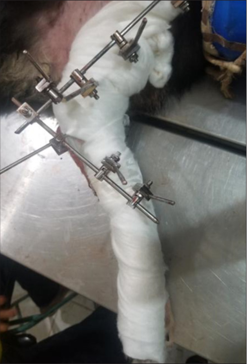
- A modified Robert Jones bandage was applied to protect the limb and control postoperative swelling.
| Parameters | Day 1 | Day 2 | Day 3 | Day 4 | Day 5 | Reference |
|---|---|---|---|---|---|---|
| Temperature (°C) | 38.8 | 38.7 | 39.5 | 39.4 | 37.8 | 38.5–39.4 |
| Pulse rate (beats/min) | 112 | --- | 102 | 126 | 130 | 90–120 |
| Respiratory rate (cycles/minutes) | 28 | --- | 30 | 2 | 26 | 20–30 |
Keys: -----(unremarkable)
Three months after the surgery, the patient was bearing substantial weight on the injured limb with the absence of pain at the luxated elbow when palpated and manipulated. However, the transfixion pins in the humerus had considerably loosened, while those in the radius were still firm. A craniocaudal radiographic evaluation showed an aligned and healed elbow joint [Figure 6]. Hence, the fixator was removed. The pin holes were cleaned, dressed, and allowed to heal at home. The patient became lame post removal of the fixator for 2 days but later gradually started bearing weight on the limb. One-year post-fixator removal, the patient was re-presented to the clinic for antirabies vaccination and was doing great on the operated limb. The patient could walk without any noticeable lameness, but lameness became obvious when the patient ran.

- A craniocaudal radiographic evaluation at two and a half months post-surgery showed an aligned and healed elbow joint.
DISCUSSION
Management of cubital re-luxation, especially from congenital elbow luxation or road traffic accidents in small animals, is not commonly reported, most likely because surgical options are often challenging but evolving. Delayed intervention in cubital re-luxation worsens prognosis, leading to joint fibrosis, muscle contracture, and osteoarthritis, making reduction difficult and often requiring salvage procedures like arthrodesis. In this case, the right elbow exhibited complete lateral radioulnar luxation, which was initially addressed with cerclage wire, yet experiencing a relapse in luxation. Employing an ESF for managing cubital re-luxation post-initial surgery proved highly efficacious.
Management of congenital luxation might prove difficult. However, in this scenario, the absence of certain congenital elbow luxation-related anomalies, such as hypoplasia or aplasia of the medial coronoid and anconeal processes Decamp et al.[3] including the history of the patient, led us to classify the case as traumatic subluxation rather than congenital, highlighting the distinct traumatic nature of the condition. The distinctive clinical manifestation of cubital luxation in an animal renders diagnosis relatively straightforward, often facilitated by imaging techniques like X-rays. Typically, the radius and ulna exhibit lateral dislocation, while the position of the anconeus process may vary, sometimes resting on the posterolateral aspect of the lateral epicondyle. It is important to note that surgically treated elbow luxation and subluxation usually develop secondary degenerative joint disease[11,12] such as bone spur and osteoarthritis, especially in older dogs, and this might be the reason why veterinarians do not always consider managing it the second time. The pain, cost, and instability of not managing the condition might cause significant inconvenience to the patient and client themselves and affect the patient’s overall quality of life of the affected dog.
Reduction of an uncomplicated luxation usually can be effected merely by manipulation[10] depending on the nature of the condition presented and sometimes use of external fixators can be applied to radius-ulna bone conditions of all ages.[13] However, in this present case, the patient who was involved in a traffic accident was supported with external skeletal fixation to help the patient obtain more stability and improve her overall quality of life after the management. According to Rahal et al.,[14] if the two bones are not held in their proper positions, the joint may easily slide as soon as the dog puts weight on the affected limb, and this might be the reason why the initial management with cerclage wire was not effective for this patient.
ESFs are frequently employed in veterinary orthopedics for stabilizing fractures and come in various configurations. They serve as both primary fixation and supplementary stabilization for a diverse range of conditions, rendering them highly adaptable and versatile instruments in veterinary practice[15] and providing more stability than the use of. Even in cases where the trans articular pin was temporarily used to manage elbow luxation, there was persistent subluxation of the humeroulnar and humeroradial joints despite the patient having adequate elbow function, even though the goal of surgical repair is not only to reconstruct the joint entirely but also to maintain the normal function of the affected limb. Several studies have highlighted potential complications associated with the use of transarticular pins in managing joint luxations. Rahal et al., Withrow, and Clark et al [14,16,17]. have reported concerns such as disruption of joint healing phases, implant failure, and the risk of bacterial migration along the implant tract. Furthermore, prolonged use of external coaptation devices has been associated with secondary complications, including articular cartilage trauma and impaired limb development, particularly in growing animals. However, the advantage of using the ESFs over the use of a transarticular pin is to provide limb stability and prevent future sub-luxation. As stated by Ozsov and Altunatimaz,[18] the early onset of weight-bearing is a significant advancement aimed at preventing potential complications such as joint stiffness and the degeneration of bones and muscles after the management. This enables a prompt restoration of limb function, thus promoting overall recovery. The use of ESF also aids animals in putting weight on the joint gradually until the joint is healed. Nnaji et al.[19] also noted that the early weight bearing of the dogs is seen when external fixatives are used and provide good joint mobility within the first 24 h post-operative. The observed early weight-bearing might be explained by the effective stabilization and improved capacity for distributing loads of the fixation devices.
As proved earlier by Milton et al. and Putnam and Archibald[11,12], surgically treated elbows luxation and subluxation usually develop secondary degenerative joint disease. However, in this particular case, there were no sign of degenerative changes observed after 1 year of post-surgery, which makes us recommend this technique for clinicians to use and avoid the fear noted earlier. We conclude that the use of a transarticular external skeletal fixation was effective for the treatment of cubital re-luxation.
However, some of the major limitations of transarticular external skeletal fixation in managing fractures in dogs are joint stiffness and reduced range of motion due to prolonged immobilization, which we also noted in our case. In addition, complications such as pin loosening, pin tract infections, and difficulty in achieving perfect alignment may affect healing and long-term limb function.[20,21] This report has a particular limitation, as it presents a subjective evaluation of a single case (i.e., not a case series).
CONCLUSION
Cubital joint re-luxation is a challenging orthopedic condition in dogs that requires prompt diagnosis and appropriate surgical intervention for optimal outcomes. In this clinical report, we present the management of cubital joint re-luxation in a 6-month-old Alsatian cross-female puppy, emphasizing the use of transarticular ESF in achieving successful surgical management.
Ethical approval:
Institutional Review Board approval is not required.
Declaration of patient consent:
Patient’s consent is not required as there are no patients in this study.
Conflicts of interest:
There are no conflicts of interest.
Use of artificial intelligence (AI)-assisted technology for manuscript preparation:
The authors confirm that there was no use of artificial intelligence (AI)-assisted technology for assisting in the writing or editing of the manuscript and no images were manipulated using AI.
Financial support and sponsorship: Nil.
References
- Idiopathic Anterior Dislocation of the Radial Head: Symptoms, Radiographic Findings, and Management of 8 Patients. J Shoulder Elbow Surg. 2019;28:1468-75.
- [CrossRef] [PubMed] [Google Scholar]
- The Elbow Joint In: Handbook of Small Animal Orthopedics and Fracture Repair (5th ed). Amsterdam, the Netherlands: Elsevier; 2016. p. :327-65.
- [CrossRef] [Google Scholar]
- The Radiological Features of Congenital Elbow Luxation/Subluxation in the Dog. J Small Anim Pract. 1982;23:621-30.
- [CrossRef] [Google Scholar]
- Stifle Joint Arthrodesis for Treating Chronic-Osteoarthritis-Affected Dogs. Vet Sci. 2023;10:407.
- [CrossRef] [PubMed] [Google Scholar]
- Correlation of Arthrodesis Stability with Degree of Joint Fusion on MDCT. AJR Am J Roentgenol. 2009;192:496-9.
- [CrossRef] [PubMed] [Google Scholar]
- Diseases of joints and Ligaments In: Tilley LP, Smith FW, eds. Handbook of Small Animal Practice (5th ed). St. Louis, MO: Elsevier; 2008. p. :763-77.
- [CrossRef] [Google Scholar]
- Traumatic Luxation of the Cubital Joint (Elbow) in Dogs: 44 Cases (1978-1988) J Am Vet Med Assoc. 1992;201:1760-5.
- [CrossRef] [PubMed] [Google Scholar]
- Veterinary Clinics of North America: Small Animal Practice. Vol 1. W.B. Saunders: United States; 1971. p. :429-40.
- [Google Scholar]
- Congenital Elbow Luxation in the Dog. J Am Vet Med Assoc. 1979;175:572-82.
- [CrossRef] [PubMed] [Google Scholar]
- The Stader Reduction Splint for Treating Fractures of the Shafts of the Long Bones 1942. Clin Orthop Relat Res. 1993;293:3-7.
- [CrossRef] [Google Scholar]
- Reduction of Humeroulnar Congenital Elbow Luxation in 8 Dogs by Using the Transarticular pin. Can Vet J. 2000;41:849-53.
- [Google Scholar]
- Postoperative Complications Associated with External Skeletal Fixators in Dogs. Vet Comp Orthop Traumatol. 2018;31:137-43.
- [CrossRef] [PubMed] [Google Scholar]
- Management of Congenital Elbow Luxation by Temporary Transarticular Pinning. Vet Med Small Anim Clin. 1977;72:1597-602.
- [Google Scholar]
- Surgical Management of Suspected Congenital Luxation of the Radial Head in Three Dogs. N Z Vet J. 2010;58:103-9.
- [CrossRef] [PubMed] [Google Scholar]
- Treatment of Extremity Fractures in Dogs Using External Fixators with Closed Reduction and Limited Open Approach. Vet Med. 2003;48:133-40.
- [CrossRef] [Google Scholar]
- Osteomedullography of Tibial Fractures Immobilized with Different Fixators in Mongrel Dogs. Anim Sci Rep. 2015;9:114.
- [Google Scholar]
- Fractures of the Tibia and Fibula In: Johnston SA, Tobias KM, eds. Veterinary Surgery: Small Animal (2nd ed). Philadelphia, PA: Elsevier Saunders; 2018. p. :1176-93.
- [Google Scholar]
- Linear External Skeletal Fixation Applied in Minimally Invasive Fashion for Stabilization of Nonarticular Tibial Fractures in Dogs and Cats. Vet Surg. 2023;52:249-56.
- [CrossRef] [PubMed] [Google Scholar]


