
Translate this page into:
Management of Hepatozoonosis and Helminthosis in Python sebae

*Corresponding author: Enokela Shaibu Idoga, Department of Veterinary Physiology, Biochemistry and Pharmacology, University of Jos, Jos, Nigeria. danooavaz@gmail.com
-
Received: ,
Accepted: ,
How to cite this article: Idoga ES, Avazi DO, Okonkwo AR, Ochai P, Dasplang KP, Uchendu C, et al. Management of Hepatozoonosis and Helminthosis in Python sebae. Res Vet Sci Med. 2025;5:4. doi: 10.25259/RVSM_13_2024
Abstract
Four African rock pythons at the Wildlife Park in Nigeria were reported to show clinical signs of reduced appetite and lethargy. In response, the management of the University of Jos Veterinary Teaching Hospital assembled a team to investigate and manage the condition. Initially suspecting helminthosis, the team decided to deworm the snakes using Levamisole at a dose of 10 mg/kg subcutaneously (SC). Whole blood, ectoparasite, and fecal samples were collected. During the handling of the snakes, they were observed to be heavily infested with ticks. Laboratory results later showed the presence of Hepatozoon spp. in the blood and nematode eggs in the feces, prompting a follow-up visit 2 weeks later. During the second visit, the snakes were treated with an injection of ivermectin at 0.1 mg/kg SC, imidocarb dipropionate at 5 mg/kg SC, and atropine sulfate at 0.02 mg/kg SC. Following treatment, the snakes made a full recovery and significantly regained their activity and appetite.
Keywords
Hepatozoon
Nematode eggs
Python sebae
Ticks

INTRODUCTION
Python sebae, commonly known as the African rock python, can be found all over sub-Saharan Africa; however, they tend to avoid the driest deserts and mountain heights with the lowest temperatures. They are among the longest snake species globally, with lengths of up to 6 m (10–16 ft).[1]
Once abundant across various regions, Python sebae, are now predominantly confined to specific habitats. These magnificent serpents have retreated to hunting reserves, national parks, and remote stretches of the African savannah, where they can thrive away from human encroachment. In these secluded environments, they can hunt their natural prey and maintain their essential role in the ecosystem.[2]
Hepatozoon is a genus of protozoa classified within the phylum Apicomplexa, which includes various parasitic organisms. Members of this genus are known to infect a range of hosts, primarily vertebrates.[3] Ticks play a crucial role as vectors for Hepatozoon, facilitating the transmission of these protozoan parasites from one host to another. This vector-host relationship is significant in the life cycle of Hepatozoon, as the parasites often rely on these blood-feeding parasites to spread their infection and maintain their population in the wild. Understanding the dynamics between Hepatozoon and its tick vectors is essential for developing strategies to control the spread of these protozoan infection.[3] Hepatozoon species are the most prevalent intracellular protozoan parasites that infect snakes, residing within their host cells and often leading to various health issues.[4,5] These parasites can have significant impacts on the overall well-being and survival of snake populations.[6] Typical clinical manifestations of hepatozoonosis in vertebrates encompass lethargy, anorexia, regurgitation, dyspnea, anemia, muscle atrophy or discomfort, and occasionally, dermal lesions.[4]
Many Hepatozoon species are non-pathogenic because they adapt effectively to their hosts. During asexual reproduction, liver, lung, kidney, and spleen cells might be damaged. Moderate-to-severe hepatozoonosis symptoms include anemia, cachexia, fever, lethargy, hyperglobulinemia, weight loss, and anorexia. This syndrome affects mammals, reptiles, birds, and amphibians.[4,7]
CASE REPORT
History and clinical examination
Officials at the Jos Wildlife Park requested the Veterinary Teaching Hospital at the University of Jos to provide care for four captive African Rock Pythons in their facility. The snakes were said to be anorexic, exhibiting a reluctance to ingest their monthly diet primarily consisting of live rabbits. The wildlife attendants indicated that a snake had killed a rabbit without ingesting it. They also reported decreased activity levels and responsiveness. The snakes had never received medical care in their 4 years of inhabiting this facility, before this call, and the sexes of the snakes were also not known.
Handling and physical examination
First visit
With initial suspicion of helminthiasis, following reported clinical signs and medical history, a team was set up to attend to these creatures. Each snake was captured and restrained physically using snake hooks and tongs, with absolute care to avoid injury to them and members of the team. The team conducted a physical examination of each snake from head to tail while the snakes were restrained. After this examination, they collected oral swabs, ectoparasite samples, and fecal samples for laboratory analysis. Blood samples were also drawn from the caudal vein and placed in heparinized sample bottles for hematology and hemoparasite screening. Following these procedures, each snake was weighed using a bathroom scale. To do this, the handler was weighed first, and then, the weight was measured again while carrying the snake. The snake’s weight was then calculated by subtracting the weight of the handler from his weight while carrying the snake. The snakes were then treated for helminthiasis (routine deworming) with the plan to return after receiving laboratory test results. To ensure team and snake safety, each snake was placed in snake bags after treatment and only released once all other snakes had been handled.
Clinical signs
Clinical signs observed include anorexia, lethargy, and heavy tick infestation.
Initial treatment
On the first visit, the snakes were treated with levamisole, at 10 mg/kg subcutaneously (SC).
Laboratory test results
Laboratory tests were carried out at the pathology, parasitology, and entomology laboratories of the University of Jos Veterinary Teaching Hospital, University of Jos, Jos, Nigeria. The laboratory results revealed the tick samples to be of the Amblyomma genus. Fecal floatation test showed presence of nematode eggs, and Hepatozoon parasites were observed in a thin blood smear of all of the snakes’ blood [Figures 1-3].

- Four (4) African rock pythons (Python sebae) in their abode at the Jos Wildlife Park.
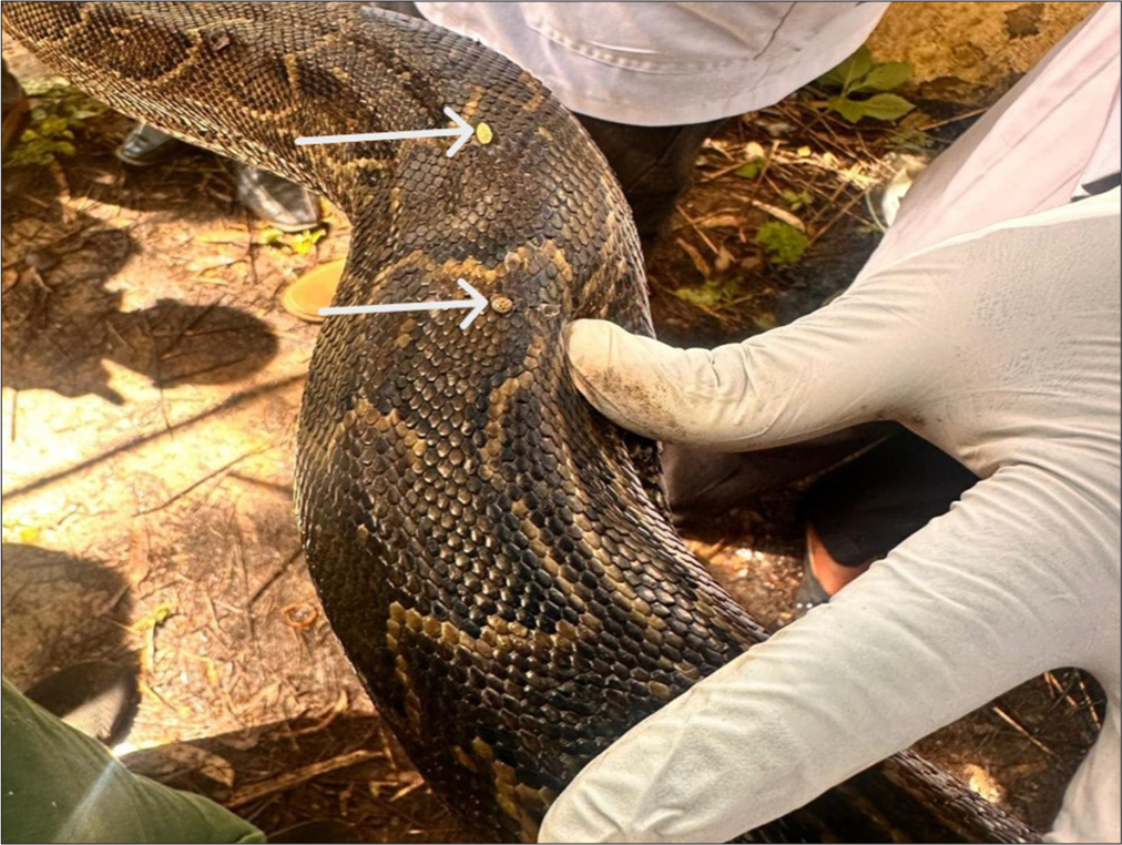
- African rock python (Python sebae) showing ticks (white arrows) on the skin.
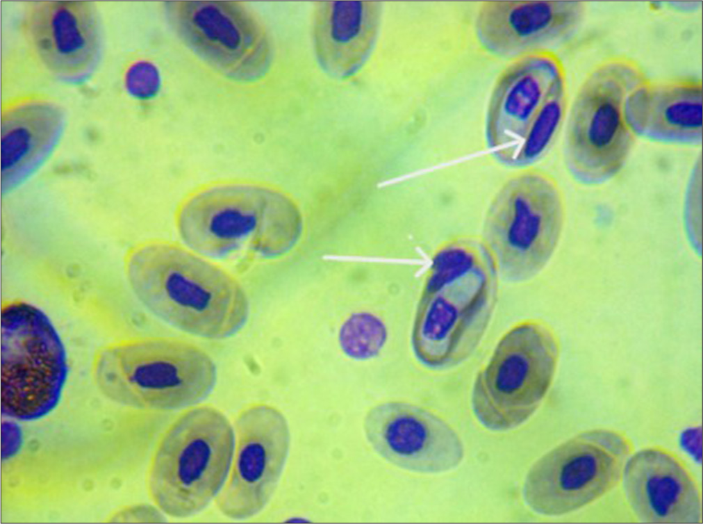
- Photomicrograph of thin blood smear of African rock python (Python sebae) showing intraerythrocytic Hepatozoon spp. (white arrows).
Second visit
The second visit to the snakes occurred 2 weeks after the first. During this visit, handling and physical examination were carried out as described earlier. The snakes were treated using 1% ivermectin (Interchemie werken “De Adelaar” B. V. Holland) at 0.1 mg/kg SC, once, imidocarb dipropionate (Interchemie werken “De Adelaar” B. V. Holland) at 5 mg/kg SC and atropine sulfate at 0.02 mg/kg SC. A summary of medications used and the quantity administered is as shown in Table 1.
| S/No. | Snakes | Weight (Kg) | Quantity of drugs administered (mg) | |||
|---|---|---|---|---|---|---|
| Levamisole 10 mg/kg SC | Ivermectin 0.1 mg/kg SC | Imidocarb 5 mg/kg SC | Atropine 0.02 mg/kg SC | |||
| 1. | Snake 1 | 9.7 | 97 | 0.97 | 48.5 | 0.194 |
| 2. | Snake 2 | 12.3 | 123 | 1.23 | 61.5 | 0.246 |
| 3. | Snake 3 | 24 | 240 | 2.4 | 120 | 0.48 |
| 4. | Snake 4 | 19.5 | 195 | 1.95 | 97.5 | 0.39 |
SC: Subcutaneously
Outcome
The snakes were said to exhibit increased activity and a significantly enhanced appetite. No evident indications of medication toxicity were observed.
DISCUSSION
Hepatozoon spp. is recognized as the most prevalent intracellular protozoan that affects snake populations.[4] To date, researchers have identified over 300 distinct species of Hepatozoon in various vertebrate hosts, with more than 120 species specifically known to infect snakes.[8] Despite this diversity, significant gaps exist in our understanding of how these parasites impact their reptilian hosts. There is a notable lack of comprehensive research information addressing the clinical signs that may arise due to Hepatozoon infection in snakes, as well as the associated pathological changes observed in affected individuals. This deficiency in knowledge hinders efforts to effectively diagnose, treat, and manage Hepatozoon infections in snakes, underscoring the need for further studies in this area.
While the specific species of Hepatozoon involved in this case remains unidentified, research has shown that certain Hepatozoon species exhibit a remarkable lack of host specificity. This means that they can infect a diverse range of host organisms, even those that are not closely related. In fact, several Hepatozoon species have been documented across a wide array of vertebrate groups, indicating their ability to utilize various definitive and intermediate hosts with minimal restrictions. This adaptability highlights the ecological versatility of Hepatozoon and raises important considerations for understanding host-parasite interactions and potential disease transmission in wildlife and domestic animals.[9]
Research on hepatozoon infections has yielded mixed results across various vertebrate taxa, including lizards, snakes, and birds. Some studies indicate that these infections produce little to no noticeable effects or only minor issues. However, other investigations reveal a much darker picture, highlighting severe health consequences. Affected animals may experience a range of symptoms, including increased white blood cell counts (leukocytosis), significant loss of appetite (anorexia), muscle damage (myopathy), and in some cases, these infections can even prove fatal.[10] The clinical signs associated with this case, anorexia and lethargy, are consistent with those reported by Wozniak et al.[11] in lizards. Despite Hepatozoon species being considered non-pathogenic due to their superior host adaptation, certain cells (liver, lung, kidney, and spleen) may nonetheless sustain damage during their asexual reproduction.[4] Hepatozoonosis is characterized by moderate–to-severe symptoms, including anemia, cachexia, fever, lethargy, hyperglobulinemia, weight loss, and anorexia, across a wide range of animals, including mammals, reptiles, birds, and amphibians.[4,7]
Tick infestation observed in this case is a common finding on snakes and underscores the importance of ticks as vectors of this parasite. Natusch et al.[12] reported the presence of ixodid ticks on snakes in Australia. The use of levamisole at 10 mg/kg for deworming the snakes is in agreement with the recommendation of De la Navarre[13] who reported that levamisole at 5–10 mg/kg Per Os or SC in snakes was effective and safe in the treatment of nematode infections in snakes. Ivermectin is one of the less toxic antiparasitic medications used to treat ectoparasite infestations in snakes.[14] Ivermectin was used in this case to tackle tick infestation and not for its antinematodal effects. Imidocarb dipropionate is the primary therapeutic agent for the treatment of Hepatozoon infections, including those in snakes. Administering the medication at a dosage of 5–6 mg/kg either SC or intramuscular injection is deemed safe and effective for the treatment of hepatozoonosis in snakes.[15] The chosen dosage of 5 mg/kg in this instance was intended to mitigate the potential of toxicity. Furthermore, atropine sulfate is employed to mitigate the secondary muscarinic effects linked to imidocarb.
CONCLUSION
Tick and helminth infestations pose significant challenges for Python sebae in captivity. Hepatozoon spp may be transmitted by these ticks, resulting in clinical manifestations such as lethargy and diminished appetite, particularly in cases of concurrent helminthosis. Treatment with Ivermectin (0.1 mg/kg SC), Imidocarb diproprionate (5 mg/kg SC), and Atropine sulphate (0.02 mg/kg SC) was both efficacious and safe for addressing these ailments. Levamisole (10 mg/kg SC) was also effectively utilised to treat helminthosis in Python sebae.
Acknowledgments
The author appreciates the immense contributions of Mr. James G and Mr. Abdulkareem YI for assisting with the laboratory analysis.
Ethical approval
The Institutional Review Board approval is not required.
Declaration of patient consent
Patient’s consent not required as there are no patients in this study.
Conflicts of interest
There are no conflicts of interest.
Use of artificial intelligence (AI)-assisted technology for manuscript preparation
The authors confirm that there was no use of artificial intelligence (AI)-assisted technology for assisting in the writing or editing of the manuscript and no images were manipulated using AI.
Financial support and sponsorship: Nil.
References
- Dark Florida: Animal Attacks, Historic Murders, Deadly Disasters and other Calamities Arcadia Publishing; 2023. p. :144.
- [Google Scholar]
- Hematological and Plasma Chemistry Values for the African Rock Python (Python sebae) Int J Vet Sci Med. 2017;5:181-6.
- [CrossRef] [PubMed] [Google Scholar]
- Molecular Identification and Clinicopathological Findings of Hepatozoon sp. Infection in a Cat: First Report from Turkey. Türk Parazitol Derg. 2018;42:286.
- [CrossRef] [PubMed] [Google Scholar]
- Discovery of a New Hepatozoon Species Namely Hepatozoonviperoi sp. nov. in Nose-horned Vipers in Türkiye. Sci Rep. 2023;13:9677.
- [CrossRef] [PubMed] [Google Scholar]
- Morphologic and Morphometric Analysis of Hepatozoon spp. (Apicomplexa Hepatozoidae) of Snakes. Mem Inst Oswaldo Cruz. 2002;97:1169-76.
- [CrossRef] [PubMed] [Google Scholar]
- Phylogenetic Relationship of Hepatozoon Blood Parasites Found in Snakes from Africa, America and Asia. Parasitology. 2014;141:389-98.
- [CrossRef] [PubMed] [Google Scholar]
- Hepatozoonosis. In: In Arthropod-borne Infectious Diseases of the Dog and Cat. United States: CRC Press; 2016. p. :121-36.
- [CrossRef] [Google Scholar]
- Hepatozoonosis In: Greene's Infectious Diseases of the Dog and Cat. United States: WB Saunders; 2021. p. :1230-47.
- [CrossRef] [Google Scholar]
- The Biology and Identification of the Coccidia (Apicomplexa) of Carnivores of the World London: Academic Press; 2018. p. :712.
- [Google Scholar]
- Old Pythons Stay Fit; Effects of Haematozoan Infections on Life History Traits of a Large Tropical Predator. Oecologia. 2005;142:407-12.
- [CrossRef] [PubMed] [Google Scholar]
- Characterization of the Clinical and Anatomical Pathological Changes Associated with Hepatozoon mocassini Infections in Unnatural Reptilian Hosts. Int J Parasitol. 2018;26:141-6.
- [CrossRef] [PubMed] [Google Scholar]
- Ticks on Snakes: The Ecological Correlates of Ectoparasite Infection in Free-ranging Snakes in Tropical Australia. Aust Ecol. 2018;43:534-46.
- [CrossRef] [Google Scholar]
- Common Parasitic Diseases of Reptiles and Amphibians In: CVC in San Diego Proceedings. 2008. p. :7. Available from: https://veterinarycalendar.dvm360.com/common-parasitic-diseases-reptiles-amphibians-proceedings [Last accessed on 2024 Nov 05]
- [Google Scholar]
- Deworming Protocols of Island Snakes Kept in Capitivity at the Butantan Institute. Arch Vet Sci. 2018;23:35-42.
- [CrossRef] [Google Scholar]
- Hemoparasite in Anaconda species Eunestes murinus at Conservation Center Wildlife Ilha Solteira-SP. Case Report. Rev Brasil Higien Sanid Anim. 2014;8:72-8.
- [CrossRef] [Google Scholar]


