Translate this page into:
Pathological Findings in the Carcass of a Dog following Ovariohysterectomy, and Intestinal Resection and Anastomosis

*Corresponding author: Ochuko Orakpoghenor, Department of Diagnostic Pathology, Pathology Faculty, College of Veterinary Surgeons, Zaria, Nigeria. ochuko.orakpoghenor@gmail.com
-
Received: ,
Accepted: ,
How to cite this article: Orakpoghenor O, Mohammed B, Muhammed MS. Pathological Findings in the Carcass of a Dog following Ovariohysterectomy, and Intestinal Resection and Anastomosis. Res Vet Sci Med. 2024;4:2. doi: 10.25259/RVSM_5_2024
Abstract
This report presented the gross and histopathological findings in the carcass of a Nigerian Indigenous Dog (NID) following ovariohysterectomy, intestinal resection and anastomosis. The carcass of a 9-month-old NID was presented to the Necropsy Unit of the Veterinary Teaching Hospital, Ahmadu Bello University Zaria, Nigeria. History was obtained, general examination of the carcass and necropsy were conducted. History revealed sudden death 2 days post-abdominal surgical procedures (ovariohysterectomy, and small intestinal resection and anastomosis). The clinical signs observed before death were weakness and dyspnea, while mild tick infestations and bilateral congested ocular mucus membranes were on general examination of the carcass. The gross pathological findings were hemorrhagic trachea, congested and hemorrhagic lungs, enlarged heart with epicardial and endocardial hemorrhages, enlarged and congested liver, enlarged spleen, hemorrhage in the gastric mucosa, intestinal hemorrhages, and an area of hemorrhagic infarct at the intestinal anastomosed site. On histopathological examination, there was slightly thickened interalveolar walls with inflammatory cellular infiltrations, cardiac hemorrhage, and hepatic congestion. These gross and histopathological findings suggest possible systemic complications, thus, emphasizing the importance of comprehensive pre-operative assessment, and post-operative care and monitoring in veterinary surgeries.
Keywords
Surgical complications
Sudden death
Gross pathology
Histopathological findings
Nigerian indigenous dog
INTRODUCTION
Necropsy and diagnostic pathology serve as invaluable tools in veterinary medicine and offer a comprehensive postmortem analysis to elucidate the underlying causes of morbidity and mortality in animals.[1] This case report emphasizes the key role of necropsy examination and histopathological analysis in unraveling the complexities surrounding peri-and post-operative complications in veterinary surgeries. Through examination of the gross and histological findings, we aimed to unravel the pathological processes that resulted in the sudden death of a Nigerian Indigenous Dog (NID) following ovariohysterectomy and intestinal resection with anastomosis, thus, shedding light on the importance of diagnostic pathology in veterinary surgical practice.
By documenting the gross pathological changes and histopathological alterations, this report attempts to offer a deeper understanding of the underlying pathophysiological mechanisms driving peri-and post-operative complications in indigenous dog breeds. Furthermore, the insights gained from this case report may inform the development of specific peri-and postoperative management strategies and surveillance protocols, thereby enhancing patient care and outcomes in veterinary surgical settings.
CASE REPORT
The carcass of a 9-month-old NID [Figure 1a] was presented to the Necropsy Unit of the Veterinary Teaching Hospital, Ahmadu Bello University Zaria, Nigeria. The dog was reported to die suddenly 2 days after ovariohysterectomy, and small intestinal resection and anastomosis. However, the dog exhibited weakness and dyspnea, was fed with milk, administered 50% dextrose saline (intravenous), and penicillin-streptomycin injection before death.
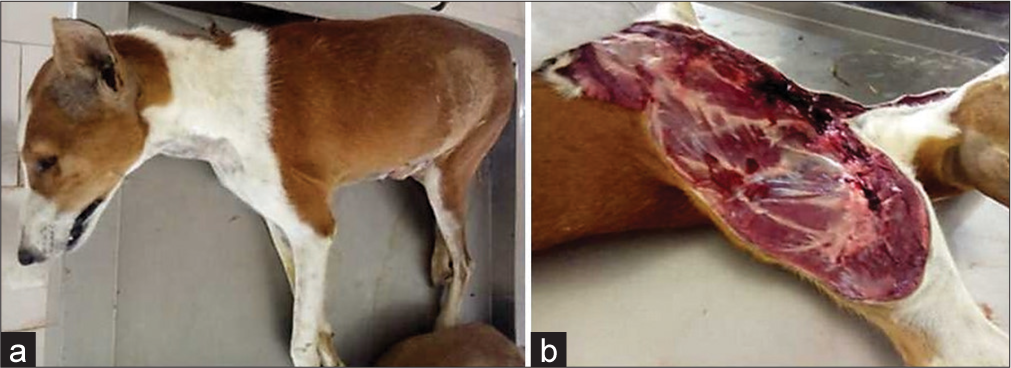
- (a) Carcass of the dog when presented, and (b) when postmortem examination commenced.
Examinations conducted
The carcass was examined and the general observations were mild tick infestation and bilateral congested ocular mucus membranes. Thereafter, a necropsy was performed [Figure 1b], and all the organs were examined for evidence of gross lesions. Sections of the lung and heart were then collected in 10% neutral buffered formalin and processed for histopathological examination.
Histopathological examination
The collected tissue sections were processed using the standard histology technique.[2] The tissues were dehydrated in progressively increasing concentrations of alcohol (70%, 80%, 90%, and absolute). Subsequently, they were cleared in xylene and embedded in paraffin wax. Following this, the tissues were sectioned at a thickness of 5 μm after being blocked and then stained with hematoxylin and eosin. The resulting slides were observed under a light microscope at various magnifications.
Gross pathological findings
The gross pathological findings were hemorrhagic trachea [Figure 2a], congested and hemorrhagic lungs [Figure 2b], enlarged heart with epicardial and endocardial hemorrhages [Figures 3a and b], enlarged and congested liver [Figure 4a], enlarged spleen [Figure 4b], hemorrhage in the gastric mucosa [Figure 5], intestinal hemorrhages, and an area of hemorrhagic infarct at the intestinal anastomosed site [Figures 6a and b].

- (a) Hemorrhagic trachea (T), (b) congested and hemorrhagic lungs (arrows).
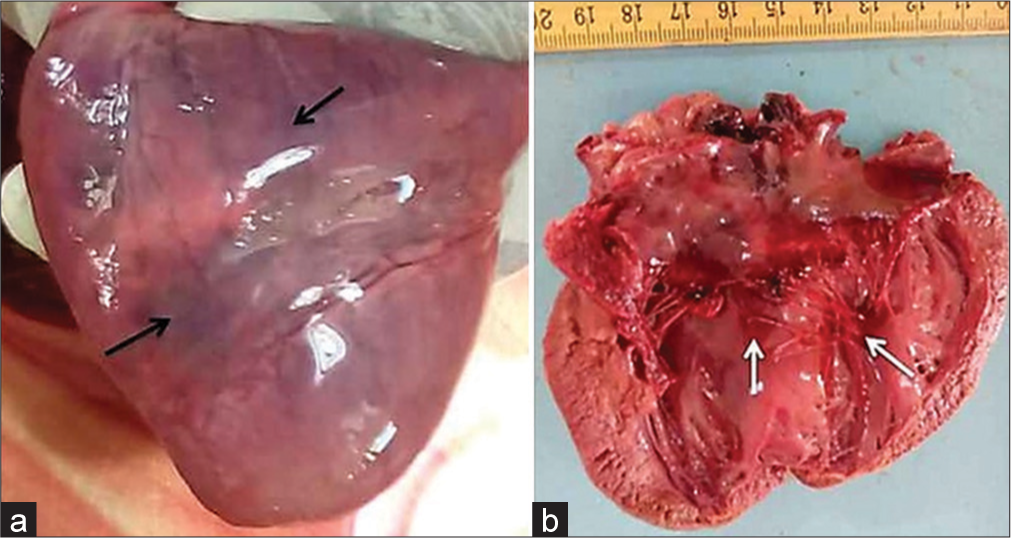
- (a) Enlarged heart with epicardial (black arrows), (b) endocardial (white arrows) hemorrhages.

- (a) Enlarged and congested liver, (b) enlarged spleen.
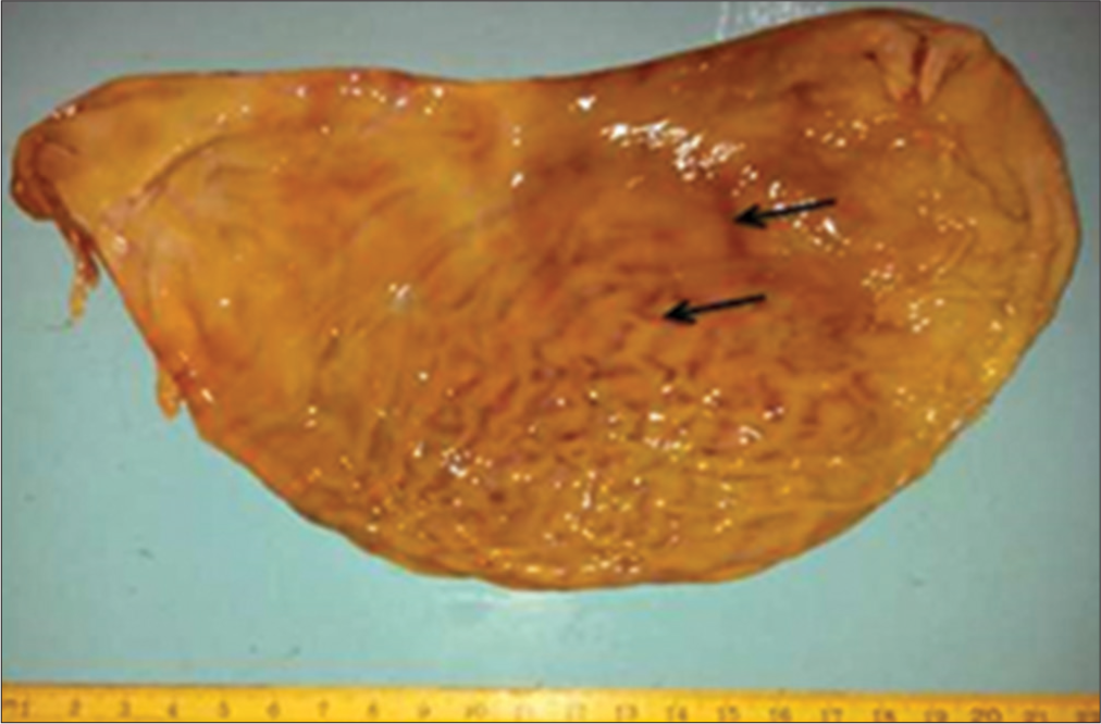
- Hemorrhages in the gastric mucosa (black arrows).
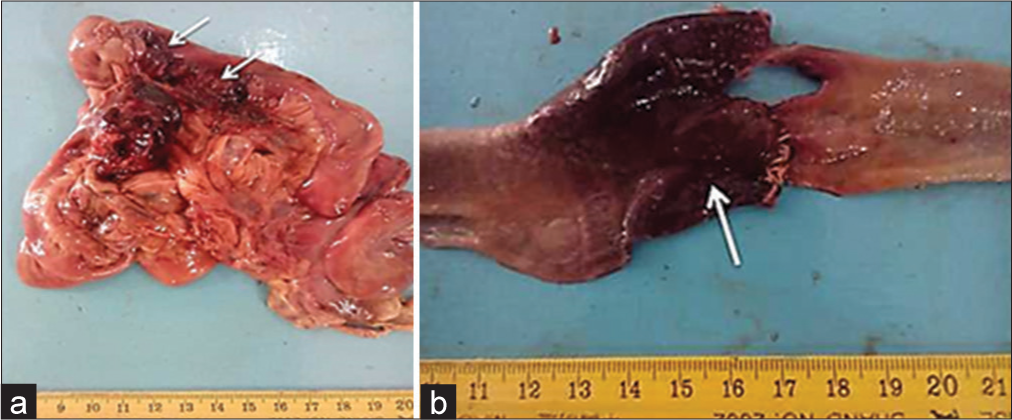
- (a) Intestinal hemorrhages (arrows), (b) an area of hemorrhagic infarct at the intestinal anastomosed site (arrow).
Histopathological findings
The histopathological findings were slightly thickened interalveolar walls and inflammatory cellular infiltrations [Figure 7a], cardiac hemorrhage [Figure 7b], and hepatic congestion [Figure 7c].
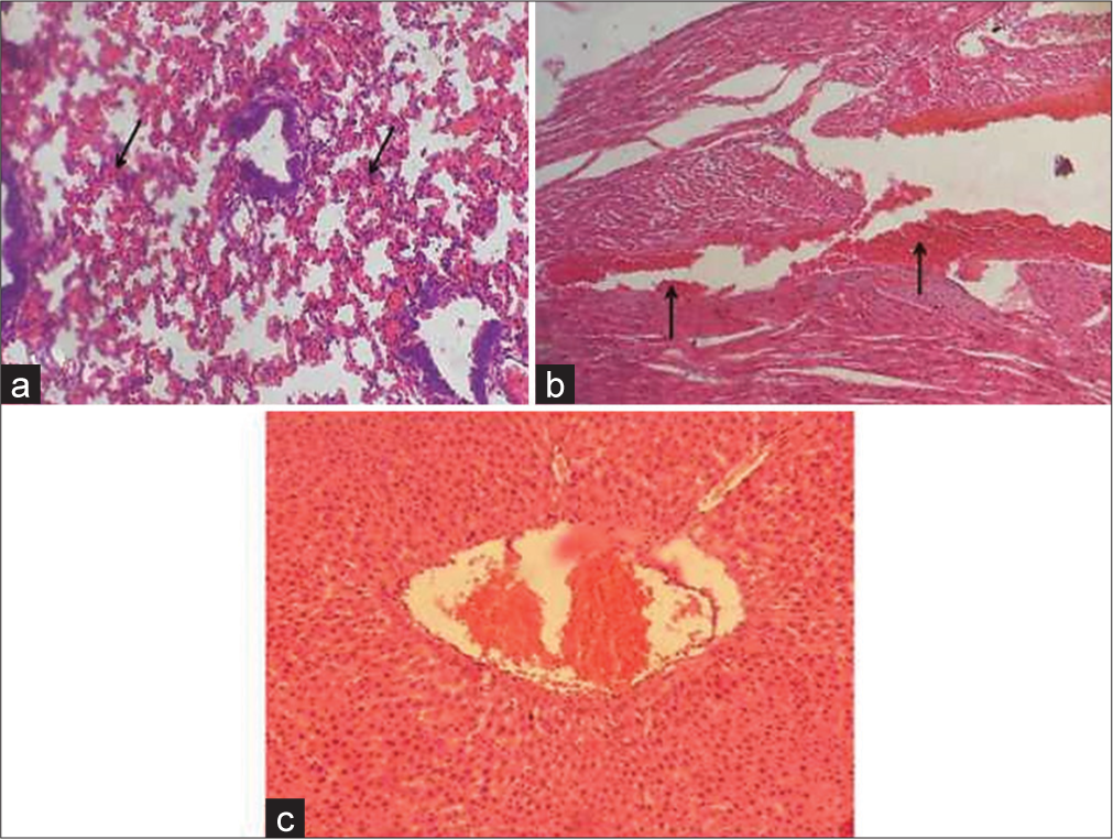
- (a) Photomicrographs of lung, (b) heart, and (c) liver sections. Note (a) slightly thickened interalveolar walls with inflammatory cellular infiltrations (arrows), (b) hemorrhages (arrows), (c) congestion hematoxylin and eosin stain ×200.
DISCUSSION
The presence of mild tick infestation and bilateral congested ocular mucous membranes in the dog in this report suggests a multifaceted approach to understanding the possible cause of death. The mild tick infestation may serve as an indicator of the overall health status of the dog and environmental exposure and can have significant impacts on health and productivity.[3] While tick infestations may not directly cause death, they can lead to complications, especially if left untreated or if the dog already has underlying health issues. Ticks can cause irritation, damage the skin, and predispose animals to bacterial and fungal infections. Tick-borne diseases can result in systemic illness, including fever, anemia, and organ dysfunction,[4] which could exacerbate the stress of surgery and compromise the ability of the dog to recover.
The bilateral congested ocular mucous membranes indicate inflammation of the ocular mucous membranes, which may reflect underlying ocular pathology or systemic conditions. Severe ocular inflammation can cause discomfort and potentially lead to complications such as corneal ulcers or secondary infections. In the perioperative period, ocular inflammation could further stress the immune system of the dog, and compromise its ability to tolerate the surgical procedures. In addition, ocular pathology can be indicative of systemic diseases or underlying conditions that may have contributed to the overall health status of the dog and prognosis.[5]
The gross and histopathological findings described in this report are indicative of severe systemic complications following the surgical procedures in the dog. These findings might suggest the development of disseminated intravascular coagulation (DIC), a life-threatening condition characterized by widespread activation of the coagulation system leading to microvascular thrombosis and subsequent organ dysfunction and hemorrhage.[6] DIC could occur as a complication of major surgical procedures, particularly those involving the gastrointestinal tract. The enlarged and congested organs, such as the heart, liver, and spleen, along with the hemorrhages in various tissues, are consistent with the consumptive and hemorrhagic phases of DIC.[7] The area of infarct at the intestinal anastomosis site further indicates compromised blood flow and tissue ischemia, which can result from the microvascular thrombosis associated with DIC.[8]
Several studies have reported similar pathological findings in dogs following ovariohysterectomy and other abdominal surgeries complicated by DIC. A study by Pearson found that 20 out of 109 dogs with complications after ovariohysterectomy had enterological problems, including vomiting, diarrhea, emaciation, and palpable abdominal masses, and these were attributed to adhesions and intestinal complications.[9] Rubio et al.[10] highlighted the changes in hematological and biochemical profiles in dogs undergoing ovariohysterectomy, showing notable changes in response to surgery, including hypoproteinemia and hypoalbuminemia. Hence, in the present case, the severe systemic involvement and the area of intestinal infarction suggest that the dog likely succumbed to the complications of DIC, which might have been triggered by the extensive surgical trauma and tissue damage from the ovariohysterectomy and intestinal resection and anastomosis procedures.
Furthermore, the hemorrhagic trachea, congested and hemorrhagic lungs, and cardiac abnormalities point toward potential respiratory and cardiovascular issues. The enlarged heart with hemorrhages could indicate cardiac stress or disease, while the hepatic congestion and splenic enlargement suggest possible circulatory disturbances. The presence of intestinal hemorrhages and hemorrhagic infarction at the anastomosed site indicate complications related to the surgical procedure. The surgical procedures might have placed significant physiological stress on the dog, potentially leading to complications such as sepsis, shock, or organ failure. The histopathological findings of thickened interalveolar walls, cardiac hemorrhage, and hepatic congestion further support the possibility of respiratory and cardiovascular compromise and circulatory disturbances, thus, contributing to the fatal outcome.
Limitations
There was no evaluation conducted for tick-borne and/or other diseases; no histopathological examinations were carried out on sections of the other organs in which gross lesions were observed.
CONCLUSION
The combination of the gross and histopathological findings could be indicative of multiple organ failure following the surgical interventions, leading to a cascade of events culminating in the sudden death of the dog. The stress of the surgeries, along with potential complications such as sepsis, shock, and organ damage, might have likely contributed to the fatal outcome in this case. Thus, an understanding of the interplay between surgical interventions, physiological responses, and pathological changes is crucial in elucidating the sequence of events that resulted in the sudden death of the dog in this study. It is recommended that there is a need for comprehensive pre-operative assessment, and post-operative care and monitoring in veterinary surgeries.
Ethical approval
Institutional Review Board approval is not required.
Declaration of patient consent
Patient’s consent not required as there are no patients in this study.
Conflicts of interest
There are no conflicts of interest.
Use of artificial intelligence (AI)-assisted technology for manuscript preparation
The authors confirm that there was no use of artificial intelligence (AI)-assisted technology for assisting in the writing or editing of the manuscript and no images were manipulated using AI.
Financial support and sponsorship
Nil.
References
- The Value of Necropsy Reports for Animal Health Surveillance. BMC Vet Res. 2018;14:191.
- [CrossRef] [PubMed] [Google Scholar]
- Manual of Histologic Staining Method of the Armed Forces Institute of Pathology (3rd ed). New York: McGraw Hill Book Company; 1968. p. :12-21.
- [Google Scholar]
- Exposure to Tick-borne Pathogens in Cats and Dogs Infested with Ixodes Scapularis in Quebec: An 8-year Surveillance Study. Front Vet Sci. 2021;8:696815.
- [CrossRef] [PubMed] [Google Scholar]
- Tick-borne Diseases of Humans and Animals in West Africa. Pathogens. 2023;12:1276.
- [CrossRef] [PubMed] [Google Scholar]
- Ocular Implications of Systemic Disease. Clin Exp Optom. 2022;105:103-4.
- [CrossRef] [PubMed] [Google Scholar]
- Early Ultrasonographic Findings after Ovariohysterectomy Operation in Bitches. Anim Health Prod Hyg. 2019;8:621-6.
- [Google Scholar]
- Disorders in Blood Circulation as a Probable Cause of Death in Dogs Infected with Babesia canis. J Vet Res. 2021;65:277-85.
- [CrossRef] [PubMed] [Google Scholar]
- Risk Factors for Dehiscence of Stapled Functional End-to-end Intestinal Anastomoses in Dogs: 53 Cases (2001-2012) Vet Surg. 2016;45:91-9.
- [CrossRef] [PubMed] [Google Scholar]
- The Complications of Ovariohysterectomy in the Bitch. J Small Anim Pract. 1973;14:257-66.
- [CrossRef] [PubMed] [Google Scholar]
- Changes in Hematological and Biochemical Profiles in Ovariohysterectomized Bitches Using an Alfaxalone-Midazolam-Morphine-Sevoflurane Protocol. Animals (Basel). 2022;12:914.
- [CrossRef] [PubMed] [Google Scholar]


