Translate this page into:
Gastroenteritis Diagnosed at Necropsy: A Review of Cases Spanning a Decade

*Corresponding author: Ochuko Orakpoghenor, Department of Veterinary Pathology, Bayero University, Kano, Kano, Nigeria. ochuko.orakpoghenor@gmail.com
-
Received: ,
Accepted: ,
How to cite this article: Muhammed MS, Hassan BA, Orakpoghenor O, Bilbonga G, Umar FS, Saleh A, et al. Gastroenteritis Diagnosed at Necropsy: A Review of Cases Spanning a Decade. Res Vet Sci Med. 2024;4:6. doi: 10.25259/RVSM_10_2024
Abstract
Objectives:
In this study, we conducted a retrospective survey of gastroenteritis diagnosed in carcasses, from 2013 to 2023, at the Necropsy Unit of the Veterinary Teaching Hospital, Ahmadu Bello University, Zaria, Nigeria.
Materials and Methods:
Data were obtained from the record book, entered into Microsoft Excel sheet, analysed using Statistical Package for Social Science (SPSS, v.27).
Results:
Results revealed that gastroenteritis was diagnosed in 20.5% of the 1052 carcasses. Yearly distribution analysis revealed fluctuations in prevalence, with peaks in 2017 (16.7%), 2018 (14.4%), and 2019 (13.0%). Species distribution analysis indicated higher proportions in canines (46.3%), caprines (31.5%), and ovines (8.3%). Sex-based analysis revealed varied proportions between males and females across different species, with no significant (p>0.05) association found between sex and species. Age distribution analysis revealed higher proportions in younger animals (<1 year) and older animals (>3 years) within specific species, and there was significant (p<0.05) association between age and species.
Conclusions:
These findings provide valuable insights into the burden of gastroenteritis in animals, thus, highlighting its temporal variations, species-, sex-, and age-specific differences. This study, therefore, contributes to the advancement of veterinary pathology, and the promotion of animal health and welfare, by improving our understanding of gastroenteritis epidemiology, and informing evidence-based veterinary practices. There is need for veterinary pathologists to develop and implement species-specific diagnostic protocols, and targeted disease management strategies to effectively mitigate the prevalence and impact of gastroenteritis in animal populations.
Keywords
Necropsy
Gastroenteritis
Pathology
Zaria
INTRODUCTION
Gastroenteritis in animals is a prevalent and multifaceted condition characterized by inflammation of the gastrointestinal tract (GIT).[1,2] It affects a wide range of species, including domestic animals such as dogs, cats, cattle, pigs, and poultry, as well as wildlife. The causes of gastroenteritis can be infectious or non-infectious.[2-4] Infectious gastroenteritis in animals is often caused by several pathogens, with bacteria such as Escherichia coli, Salmonella, Campylobacter, and Clostridium perfringens commonly implicated.[5-8] Viruses such as canine parvovirus, feline panleukopenia virus, rotavirus, and coronavirus also play significant roles, particularly in young animals.[9] Parasitic gastroenteritis (PGE) can result from infestations with protozoans such as Giardia and Cryptosporidium or helminths such as roundworms, hookworms, and tapeworms.[10] In addition, non-infectious factors such as dietary changes, ingestion of foreign bodies, toxins, stress, and underlying systemic diseases can trigger gastroenteritis reactions.[11,12]
The clinical presentations of gastroenteritis include vomiting, diarrhea, abdominal pain, anorexia, lethargy, and dehydration.[1] These clinical presentations and severity of gastroenteritis tend to vary greatly with factors such as the underlying cause, species, age, immune status, and environmental conditions.[13] The gross pathological changes associated with gastroenteritis include signs of inflammation such as redness, swelling, and increased vascularity in the affected tissues.[14,15] The mucosal lining of the GIT may appear thickened, hyperemic, and friable, with erosions, ulcers, or petechial hemorrhages observed in severe cases.[16] In addition, there may be an accumulation of mucus, fibrin, and inflammatory exudates within the lumen of the intestine.[17] On histopathology, gastroenteritis is characterized by several inflammatory lesions affecting the mucosal, submucosal, and, occasionally, the muscular layers of the GIT. These involve epithelial damage, loss of epithelial integrity, and infiltration of inflammatory cells such as neutrophils, lymphocytes, and plasma cells within the lamina propria. Edema, congestion, and hemorrhage may be observed in the submucosal layer. In chronic cases, there may be mucosal regeneration, including epithelial hyperplasia, crypt distortion, and fibrosis.[18,19]
The diagnosis of gastroenteritis involves a combination of clinical evaluation, laboratory tests, and diagnostic imaging in the live animals, and postmortem examination in the dead animals.[20] Findings from physical examinations such as abdominal pain, dehydration, and abnormal fecal consistency provide initial clues, which are further supported by laboratory analyses as well as necropsy.[21]
Necropsy, or postmortem examination, plays a crucial role in assessing the pathological changes associated with gastroenteritis in animals.[22,23] During necropsy, the GIT is examined to identify gross lesions, thus providing valuable insights into the extent and severity of gastrointestinal involvement.[24] In addition, histopathological examination of tissue samples obtained during necropsy enables the detailed characterization of inflammatory processes, which contribute to the development and progression of gastroenteritis.[25] Despite its importance, there is a notable gap in comprehensive data regarding gastroenteritis cases diagnosed at the Necropsy Unit of the Veterinary Teaching Hospital (VTH) at Ahmadu Bello University (A.B.U.) in Zaria, Kaduna State, Nigeria, from 2013 to 2023. This retrospective study seeks to address this gap by examining the prevalence, temporal trends, species, sex, and age-specific distributions associated with gastroenteritis in the local animal population.
MATERIAL AND METHODS
Location of the study
The study was conducted in the Necropsy Unit of the VTH, A.B.U. Zaria, Kaduna State, Nigeria. In this Necropsy Unit, postmortem examinations of carcasses are carried out for the purpose of establishing morphologic diagnoses and the causes of mortality.
Study design
The study was retrospective in design and involved the review of gastroenteritis diagnosed in carcasses presented to the Necropsy Unit of the VTH, A.B.U. Zaria from January 2013 to December 2023.
Data extraction
The data used for the study were obtained from the record book of the Necropsy Unit of the VTH, A.B.U. Zaria, Kaduna State, Nigeria. Gastroenteritis diagnosed in carcasses whose data were incomplete or not available was not included in this study. The data variables extracted were the year in which the gastroenteritis was diagnosed and species, sex, and age of the animal.
Data analyses
Data were assessed for completeness, entered, and cleaned in Microsoft Office Excel version 2013, and later exported to the Statistical Package for the Social Sciences (version 23.0) for analysis. Frequencies and percentages were used to express, while tables and charts were used to present the data. The associations between sex, age, and the species of animals diagnosed with gastroenteritis were tested using the Chi-square statistic. The level of significance was set at values of P ≤ 0.05.
RESULTS
Overall prevalence and yearly distribution of gastroenteritis
From 2013 to 2023, a total of 1052 carcasses were presented to the Necropsy Unit, and gastroenteritis was diagnosed in 20.5% of the carcasses [Table 1].
| Year | Total number of carcasses presented | Frequency of gastroenteritis diagnosed | Proportion (%) |
|---|---|---|---|
| 2013 | 105 | 23 | 21.9 |
| 2014 | 105 | 17 | 16.2 |
| 2015 | 193 | 19 | 9.8 |
| 2016 | 128 | 13 | 10.2 |
| 2017 | 102 | 36 | 35.3 |
| 2018 | 107 | 31 | 29.0 |
| 2019 | 128 | 28 | 21.9 |
| 2020 | 44 | 10 | 22.7 |
| 2021 | 64 | 16 | 25.0 |
| 2022 | 36 | 7 | 19.4 |
| 2023 | 40 | 16 | 40.0 |
| Overall | 1052 | 216 | 20.5 |
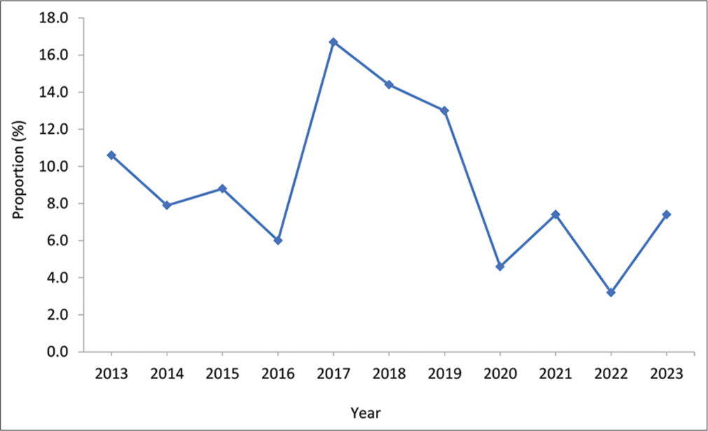
Gastroenteritis was highest (16.7%) in 2017, followed by in 2018 (14.4%) and 2019 (13.0%), and least in 2022 (3.2%) [Figure 1]. The proportions of gastroenteritis were 10.6%, 7.9%, 8.8%, and 6.0% in 2013, 2014, 2015, and 2016, respectively. In 2021 and 2023, the proportions of gastroenteritis were each 7.4%, and 4.6% in 2020 [Figure 1].

- Yearly trend of gastroenteritis diagnosed, from 2013 – 2023, at the Necropsy Unit of the Veterinary Teaching Hospital, Ahmadu Bello University Zaria.
Species, sex, and age distributions
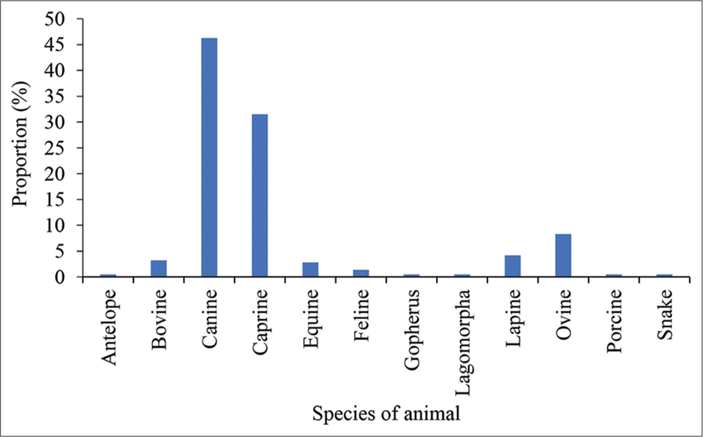
The proportion of gastroenteritis was highest (46.3%) in canine, followed by caprine (31.5%) and ovine (8.3%), and least (0.5%) in the antelope, gopherus, lagomorpha, porcine, and snake [Figure 2]. Other species of animals in which gastroenteritis was diagnosed were bovine (3.2%), equine (2.8%), feline (1.4%), and lapine (4.2%) [Figure 2].

- Distribution of gastroenteritis, based on species of animals, diagnosed from 2013 – 2023 at the Necropsy Unit of the Veterinary Teaching Hospital, Ahmadu Bello University Zaria.
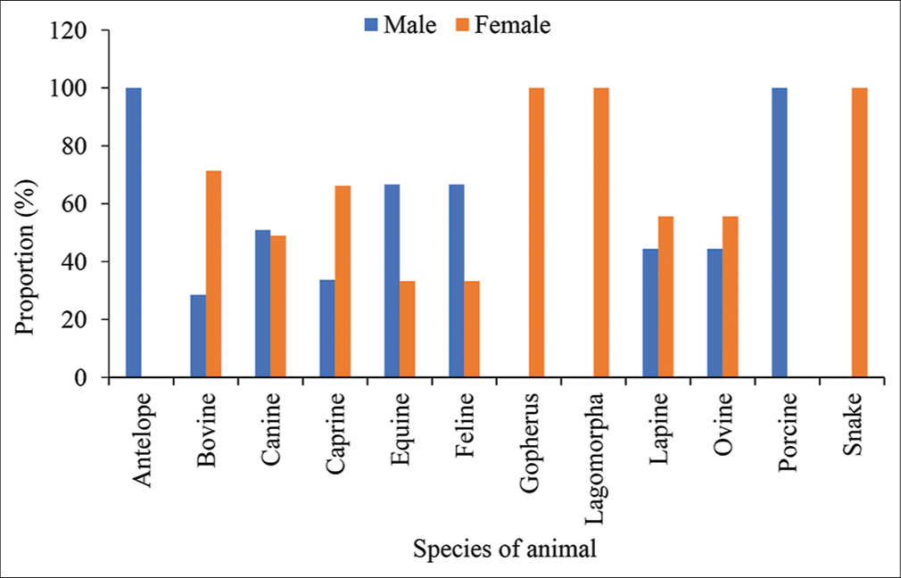
The proportion of gastroenteritis diagnosed was 100.0% in the males of antelope and porcine; and females of gopherus, lagomorpha, and snake [Figure 3]. Higher proportions of gastroenteritis were diagnosed in the males of canine (51.0%), equine (66.7%), and feline (66.7%), but in the females of bovine (71.4%), caprine (66.2%), lapine (55.6%), and ovine (55.6%) [Figure 3]. There was no significant (P > 0.05) association between the sex and species of animals in which gastroenteritis was diagnosed.

- Distribution of gastroenteritis, based on sex of animals, diagnosed from 2013 – 2023 at the Necropsy Unit of the Veterinary Teaching Hospital, Ahmadu Bello University Zaria. χ2 = 12.261, P = 0.344.
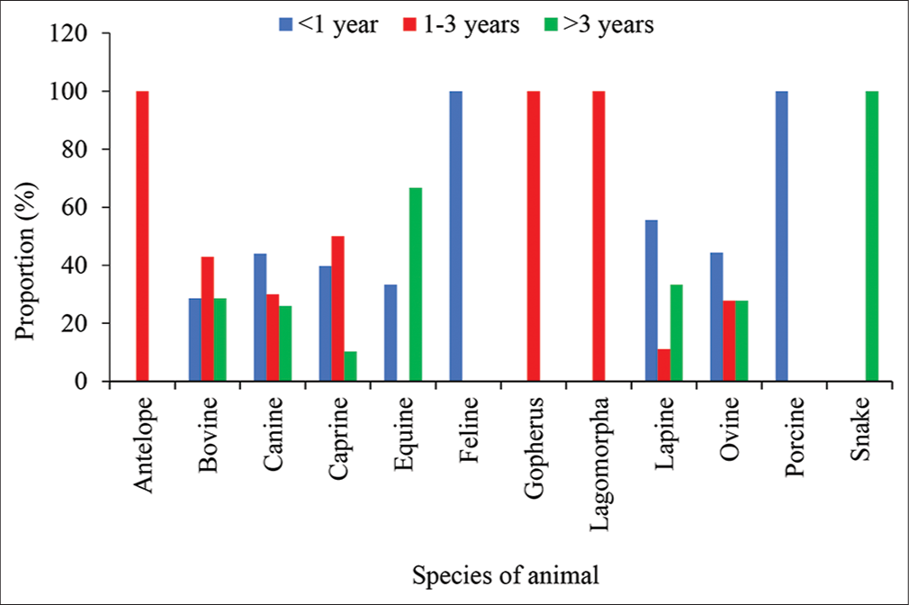
Gastroenteritis was diagnosed in 100.0% of porcine <1 year; antelope, gopherus, and lagomorpha 1–3 years; and snake >3 years old [Figure 4]. There were higher proportions of gastroenteritis in canine (44.0%), lapine (55.6%), and ovine (44.4%) <1 year; bovine (42.9%) and caprine (50.0%) 1–3 years; and in equine (66.7%) >3 years old [Figure 4]. A significant (P < 0.05) association existed between the age and species of animals diagnosed with gastroenteritis.

- Distribution of gastroenteritis, based on age of animals, diagnosed from 2013 – 2023 at the Necropsy Unit of the Veterinary Teaching Hospital, Ahmadu Bello University Zaria. χ2 = 35.614, P = 0.033.
DISCUSSION
The overall prevalence of gastroenteritis diagnosed in this study (20.5%) is consistent with the significant burden of gastrointestinal diseases reported in veterinary pathology. The previous studies have documented the widespread occurrence of gastroenteritis in animals, with varied prevalence rates depending on factors such as geographic location, species, and study design. For instance, a study by Shima et al.[26] reported a gastroenteritis prevalence of 41.2% among sick dogs following a 1-year retrospective medical records of dogs from 10 veterinary clinics in Nigeria. Kataria et al.[13] reported a 12.2% gastroenteritis prevalence in dogs in India. In a goat, severe PGE was reported.[27] In another study, the prevalence of GIT parasites reported in buffaloes and cattle was 58.59% in Pakistan.[28] These observed occurrences emphasize the importance of gastroenteritis as a common pathological condition affecting different species of animals.
The yearly distribution of gastroenteritis cases revealed temporal variations in disease prevalence over the study period, with peaks observed in certain years, such as 2017, 2018, and 2019. These findings align with previous studies that have reported seasonal and annual fluctuations in the incidence of gastrointestinal diseases in animals.[29-32] Factors such as changes in environmental conditions, management practices, and the introduction of infectious agents might have contributed to the observed fluctuations in gastroenteritis prevalence over time.[13,30,33,34] However, there is a need for further investigation into the specific factors influencing these temporal trends to enhance our understanding of disease dynamics, so as to inform targeted preventive measures.
The species distribution of gastroenteritis in this study highlighted variations in susceptibility among different animal species, with canines, caprines, and ovines identified as the most commonly affected species. This finding is consistent with previous studies that have reported species-specific differences in the prevalence of gastroenteritis.[33,35,36] The observed species distribution may reflect differences in host susceptibility, environmental exposures, and pathogen reservoirs among animal populations. Furthermore, differences in dietary habits, management practices, and genetic predispositions may also contribute to species-specific variations in gastroenteritis prevalence.[19,33,36-38]
The sex distribution of gastroenteritis cases reveals variations in disease prevalence between male and female animals within certain species. While no significant association was found between sex and species in which gastroenteritis was diagnosed, differences in prevalence between males and females were evident in specific species such as canines, equines, and bovines. These findings are consistent with studies that have reported sex-based disparities in the prevalence of gastrointestinal diseases in animals. Hormonal differences, behavioral factors, and physiological variations between male and female animals may contribute to differences in disease susceptibility and severity.[39-41]
In the present study, the age distribution of gastroenteritis showed age-related patterns in disease occurrence, with varying prevalence rates observed across different age groups. Young animals, particularly those <1 year old, exhibited higher prevalence rates of gastroenteritis compared to older age groups. Conversely, older animals, particularly those over 3 years old, also exhibited elevated prevalence rates of gastroenteritis. These findings are consistent with studies that have reported age-specific patterns in the prevalence of gastrointestinal diseases in animals.[33,42] For example, a study on the age distribution of gastroenteritis in dogs found that dogs under 1 year of age were more likely to be infected with multiple viruses compared to older dogs.[13] Factors such as immature immune systems, nutritional status, and exposure to pathogens may contribute to increased susceptibility to gastroenteritis in young animals, while age-related changes in immune function and gastrointestinal health may predispose older animals to the disease.[1] However, further research is needed to understand the complex interplay of these factors and their impact on the prevalence of gastroenteritis in different age groups of animals.
CONCLUSION
The findings of this study contribute to our understanding of the epidemiology of gastroenteritis in carcasses presented for postmortem examination at the Necropsy Unit of the VTH, A.B.U. Zaria, and highlight the complex interplay of factors influencing its occurrence and distribution. By identifying temporal trends, species-specific differences, sex-based disparities, and age-related patterns in gastroenteritis prevalence, this study provides valuable insights that can inform targeted interventions for disease prevention, management, and control.
Ethical approval
Institutional Review Board approval is not required as it is a retrospective study.
Declaration of patient consent
Patient’s consent not required as there are no patients in this study.
Conflicts of interest
There are no conflicts of interest.
Use of artificial intelligence (AI)-assisted technology for manuscript preparation
The authors confirm that there was no use of artificial intelligence (AI)-assisted technology for assisting in the writing or editing of the manuscript and no images were manipulated using AI.
Financial support and sponsorship
Nil.
References
- Norovirus Encounters in the Gut: Multifaceted Interactions and Disease Outcomes. Mucosal Immunol. 2019;12:1259-67.
- [CrossRef] [PubMed] [Google Scholar]
- Zoonotic Diseases: Etiology, Impact, and Control. Microorganisms. 2020;8:1405.
- [CrossRef] [PubMed] [Google Scholar]
- Epidemiological Investigation of Norovirus Infections in Punjab, Pakistan, through the One Health Approach. Front Public Health. 2023;11:1065105.
- [CrossRef] [PubMed] [Google Scholar]
- Enteropathogenic Escherichia coli (EPEC) Infection in Association with Acute Gastroenteritis in 7 Dogs from Saskatchewan. Can Vet J. 2016;57:964-8.
- [Google Scholar]
- Prevalence of Campylobacter Species in Human, Animal and Food of Animal Origin and their Antimicrobial susceptibility in Ethiopia: A Systematic Review and Meta-analysis. Ann Clin Microbiol Antimicrob. 2020;19:61.
- [CrossRef] [PubMed] [Google Scholar]
- Clostridium perfringens Gastroenteritis. In: Foodborne Infections and Intoxications (5th ed.). Cambridge: Academic Press; 2021. p. :89-103. Ch. 6
- [CrossRef] [Google Scholar]
- Salmonella and Salmonellosis: An Update on Public Health Implications and Control Strategies. Animals (Basel). 2023;13:3666.
- [CrossRef] [PubMed] [Google Scholar]
- Canine Parvovirus Infections and Other Viral Enteritides In: Canine and Feline Infectious Diseases. Vol 14. Netherlands: Elsevier; 2014. p. :141-51.
- [CrossRef] [Google Scholar]
- Intestinal Parasitic Infections in 2023. Gastroenterol Res. 2023;16:127-40.
- [CrossRef] [PubMed] [Google Scholar]
- Diseases of the Gastrointestinal System In: Sheep, Goat, and Cervid Medicine. Vol 21. Netherlands: Elsevier; 2021. p. :63-96.
- [CrossRef] [Google Scholar]
- A Prevalence Study on Dogs Suffering from Gastroenteritis. Pharma Innov J. 2020;9:176-9.
- [CrossRef] [Google Scholar]
- Pathomorphological and Microbiological Studies in Sheep with Special Emphasis on Gastrointestinal Tract Disorders. Vet World. 2015;8:1015-20.
- [CrossRef] [PubMed] [Google Scholar]
- Pathology of Coronavirus Infections: A Review of Lesions in Animals in the One-Health Perspective. Animals (Basel). 2020;10:2377.
- [CrossRef] [PubMed] [Google Scholar]
- Infections of the gastrointestinal tract. Diagn Pathol Infect Dis. 2018;18:232-71.
- [CrossRef] [Google Scholar]
- Infectious Diseases of the Gastrointestinal Tract In: Rebhun's Dis Dairy Cattle. Vol 18. Netherlands: Elsevier; 2018. p. :249-356.
- [CrossRef] [Google Scholar]
- Diagnostic Histopathology of Infections of the Luminal Gastro-Intestinal Tract: 'Small Blue Dots', Worms and Sexually Transmitted Infections of the Distal Gastro-intestinal Tract. Diagn Pathol. 2013;19:72-9.
- [CrossRef] [Google Scholar]
- Disorders of the Gastrointestinal System. Equine Intern Med. 2018;18:709-842.
- [CrossRef] [Google Scholar]
- Practical Guide to the Diagnostics of Ruminant Gastrointestinal Nematodes, Liver Fluke and Lungworm Infection: Interpretation and Usability of Results. Parasit Vectors. 2023;16:58.
- [CrossRef] [PubMed] [Google Scholar]
- Diagnosis and Treatment of Infectious Enteritis in Adult Ruminants. Vet Clin North Am Food Anim Pract. 2018;34:119-31.
- [CrossRef] [PubMed] [Google Scholar]
- The Value of Necropsy Reports for Animal Health Surveillance. BMC Vet Res. 2018;14:191.
- [CrossRef] [PubMed] [Google Scholar]
- Necropsy as an Important Diagnostic Step in Veterinary Pathology: The Past, Present, and Future Perspectives. Res Vet Sci Med. 2024;4:1-4.
- [CrossRef] [Google Scholar]
- Gross Lesions of Alimentary Disease in Adult Cattle. Vet Clin North Am Food Anim Pract. 2012;28:483-513.
- [CrossRef] [PubMed] [Google Scholar]
- An Endoscopic and Histopathological Assessment and Correlation of Endoscopic Score with Clinical Activity Index (CIBDAI) in the Diagnosis of Canine Idiopathic Inflammatory Bowel Disease. Iran J Vet Res. 2023;24:58-64.
- [Google Scholar]
- A Retrospective Study of the Prevalence of Gastroenteritis in Dogs Attending Some Veterinary Clinics in Nigeria. Rev Vét Clin. 2021;56:170-6.
- [CrossRef] [Google Scholar]
- Severe Parasitic Gastroenteritis (PGE) in a Goat: A Veterinary Case Report and Way Forward. Thai J Vet Med. 2019;49:295-9.
- [Google Scholar]
- Prevalence of Gastrointestinal Parasitic Infection in Cows and Buffaloes in Lower Dir, Khyber Pakhtunkhwa, Pakistan. Braz J Biol. 2023;83:e242677.
- [CrossRef] [PubMed] [Google Scholar]
- Global Patterns of Seasonal Variation in Gastrointestinal Diseases. J Postgrad Med. 2013;59:203-7.
- [CrossRef] [PubMed] [Google Scholar]
- Seasonality and Changing Prevalence of Common Canine Gastrointestinal Nematodes in the USA. Parasit Vectors. 2019;12:430.
- [CrossRef] [PubMed] [Google Scholar]
- Longitudinal Dynamics of Co-infecting Gastrointestinal Parasites in a Wild Sheep Population. Parasitology. 2022;149:1-12.
- [CrossRef] [Google Scholar]
- Seasonal Variations in Production Performance, Health Status, and Gut Microbiota of Meat Rabbit Reared in Semi-Confined Conditions. Animals (Basel). 2023;14:113.
- [CrossRef] [PubMed] [Google Scholar]
- Prevalence and Risk Factors of Gastrointestinal Parasitic Infections in Goats in Low-Input Low-output Farming Systems in Zimbabwe. Small Rumin Res. 2016;143:75-83.
- [CrossRef] [PubMed] [Google Scholar]
- Chronic Stress-Related Gastroenteric Pathology in Cheetah: Relation between Intrinsic and Extrinsic Factors. Biology (Basel). 2022;11:606.
- [CrossRef] [PubMed] [Google Scholar]
- Prevalence of Gastrointestinal Parasites in Sheep and Goats of Bui and Donga-Mantung Divisions of the North West Region of Cameroon. Asian J Animal Vet Adv. 2021;7:1-15.
- [CrossRef] [Google Scholar]
- A Comprehensive Molecular Survey of Viral Pathogens Associated with Canine Gastroenteritis. Arch Virol. 2023;168:36.
- [CrossRef] [PubMed] [Google Scholar]
- Fiber Effects in Nutrition and Gut Health in Pigs. J Anim Sci Biotechnol. 2014;5:15.
- [CrossRef] [PubMed] [Google Scholar]
- Dog-walking Behaviours affect Gastrointestinal Parasitism in Park-attending Dogs. Parasit Vectors. 2014;7:429.
- [CrossRef] [PubMed] [Google Scholar]
- Sex Differences in Immune Responses. Nat Rev Immunol. 2016;16:626-38.
- [CrossRef] [PubMed] [Google Scholar]
- Sex-Related Differences in GI Disorders. Handb Exp Pharmacol. 2017;239:177-92.
- [CrossRef] [PubMed] [Google Scholar]
- Sex-and Gender-Related Differences in Common Functional Gastroenterologic Disorders. Mayo Clin Proc. 2021;96:1071-89.
- [CrossRef] [PubMed] [Google Scholar]
- Epidemiology of Gastrointestinal Parasites of Cattle in Three Districts in Central Ethiopia. Animals (Basel). 2023;13:285.
- [CrossRef] [PubMed] [Google Scholar]


